
|
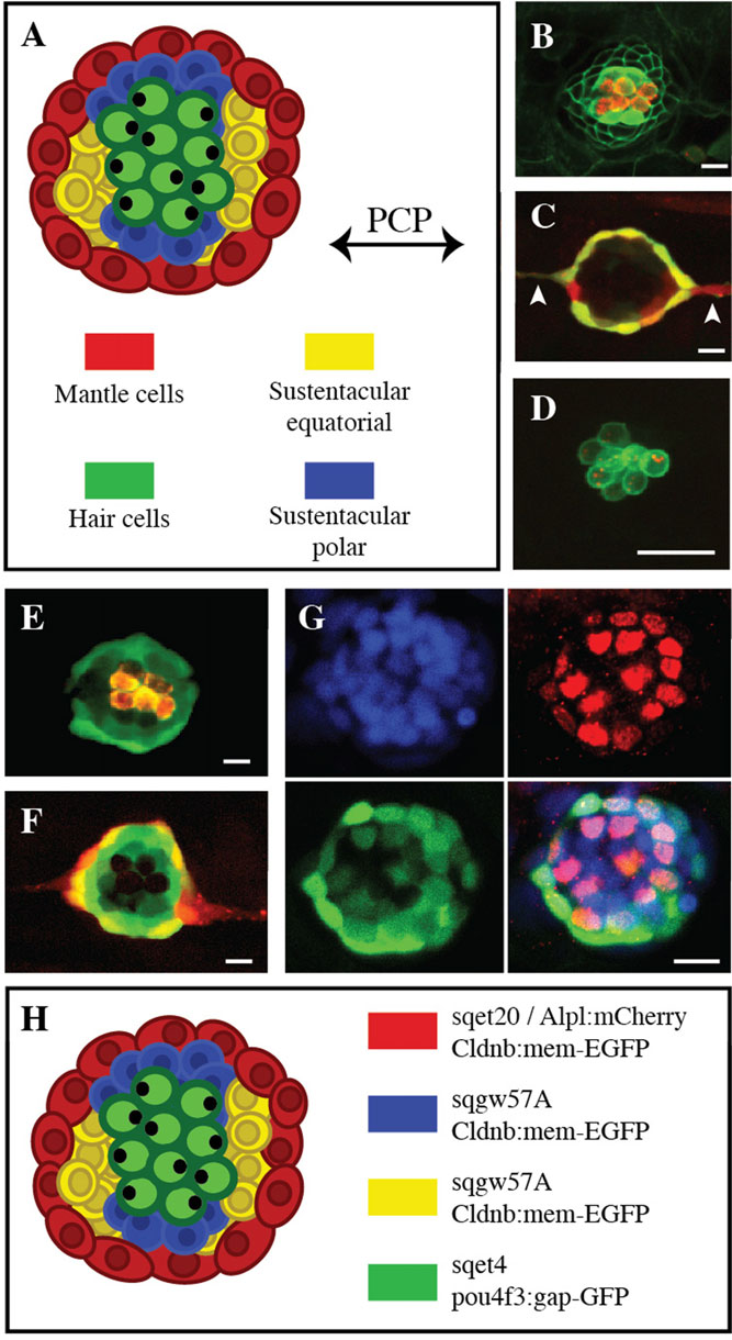
Fig. 1
Neuromast organization and transgenic zebrafish lines. (A) Scheme of a neuromast depicting different cell types and axes of symmetry. (B-F) Confocal image of a larval neuromast of (B) Tg[Cldnb:mem-EGFP;ET(krt4:EGFP)sqet4] (green), incubated in the vital dye DiAsp to reveal functional hair cells (red), (C) Tg[Alpl:mCherry (red) ; ET(krt4:EGFP)sqet20 (green)] in which white arrowhead point to interneuromast cells, (D) Tg[pou4f3:gap43-GFP (green) ; pou4f3:Ribeye-Kusabira (red)], (E) Tg[ET(krt4:EGFP)sqgw57A] (green) labeled with DiAsp (red), (F) Tg[ET(krt4:EGFP)sqgw57A (green) ; Alpl:mCherry (red)], (G) Tg[ET(krt4:EGFP)sqgw57A] (green) immunostained for Sox-2 (red) and incubated with the nuclear dye DAPI (blue). (H) Overview of a neuromast with the different cell types and transgenic markers. Scale bars=10µm.