
|
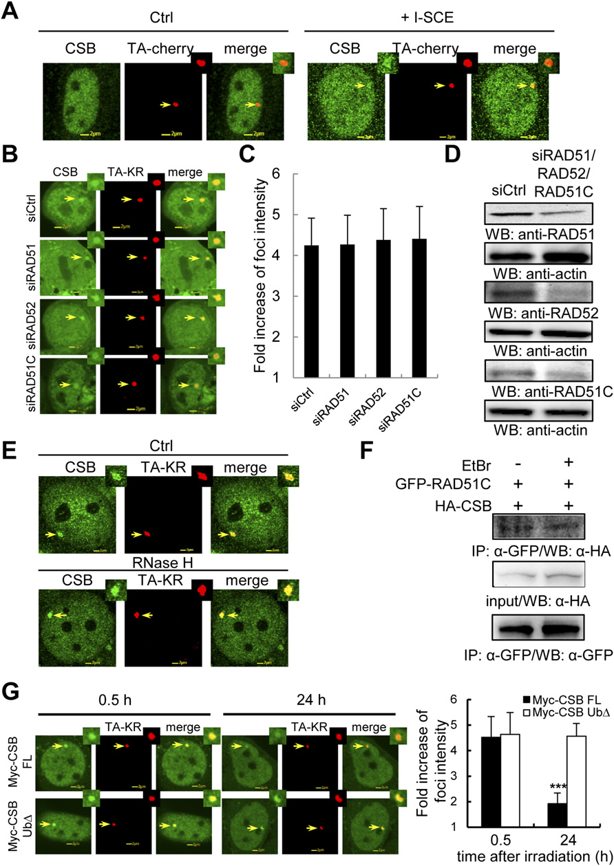
Fig. S4
CSB recruited at transcriptionally active damage sites is independent of HR factors. (A) CSB is recruited to I-SCEI induced DSBs. TA-cherry and HA-CSB with/without I-SCEI were cotransfected into U2OS TRE cells. After 24 h, cells were fixed and stained with HA-antibody. The recruitment of CSB at TA-cherry with/without I-SCEI sites is shown. (B) siRAD51, siRAD51C, or siRAD52 transfected U2OS TRE cells were expressed with FLAG-CSB and TA-KR. The recruitment of CSB at sites of TA-KR is shown after light irradiation for 10 min followed by 30 min of incubation. (C) The fold increase of CSB intensity after knockdown of the indicated protein. (D) Knockdown effects of RAD51, RAD52, and RAD51C by siRNAs. Western blot of siCtrl, siRAD52, or siRAD51C transfected U2OS TRE cells with the indicated antibody. (E) The recruitment of CSB at sites of TA-KR in U2OS TRE cells with light irradiation for 10 min followed with 30 min incubation. Cells were pretreated for 15 min with RNaseH (15 units). (F) RAD51C interacts with endogenous CSB independently of DNA after DNA damage. Flp-in 293 cells stably expressing GFP-RAD51C were treated with 5 Gy IR with 1 h postirradiation incubation. Then cell lysates with/without 10 µg/mL EtBr were applied for IP by anti-GFP. Detection of endogenous CSB is shown. (G) Myc-CSB FL or UbΔ and TA-KR cotransfected U2OS TRE cells were irradiated with light for 10 min followed by 0.5-24 h incubation. Cells were immunostained by anti-Myc. Data are represented as mean ± SD, and the P values were determined by using Student’s two-tailed t test.