
|
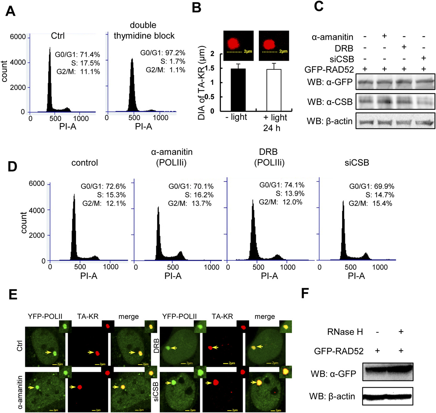
Fig. S3
Cell cycle-independent and CSB-dependent recruitment of the HR factors at TA-KR damage sites. (A) U2OS TRE cells were synchronized at the G1 phase by double thymidine block and stained with PI. The graph shows distribution of the cell population by FACS. (B) TA-KR transfected U2OS TRE cells with/without light irradiation 24 h postincubation. Diameter of TA-KR was assessed by confocal microscopy. (C–E) Western blot of GFP-RAD52 and CSB (C), cell cycle profiles (D), and recruitment of YFP-POLII (E) in U2OS TRE cells pretreated with the RNA POLII inhibitor α-amanitin (100 µg/mL) for 1 h, DRB (20 µM) for 24 h, or siCSB. D shows FACS analysis of U2OS TRE cells with the same treatment as in C. (F) Western blot of GFP-RAD52 in cells with or without RNaseH treatment (15 units) for 15 min.