
|
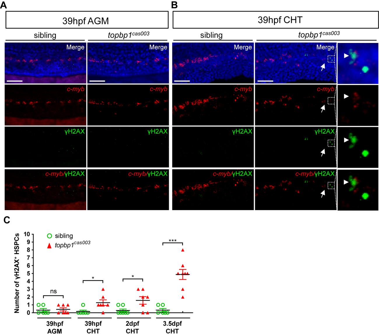
Fig. S8 DNA damage is accumulated in HSPCs in the CHT region of topbp1cas003 mutants.
(A-B) Triple staining of γH2AX antibody, c-myb fluorescent in situ hybridization and DAPI in topbp1cas003 mutants and siblings at 39hpf. The triple staining results show that the γH2AX+ HSPCs, which are undetectable in the AGM region in both mutants and siblings (A), are increased in the CHT region of topbp1cas003 mutants at 39hpf (B). The right columns in B are the magnified views of the dashed boxes in the middle columns. Scale bars represent 50um. (C) Quantification of γH2AX+ HSPCs in the AGM or CHT region in topbp1cas003 mutants and siblings at 39hpf, 2dpf and 3.5dpf. The number of γH2AX+ HSPCs is increased in the CHT region in the mutants from 39hpf to 3.5dpf. Error bars represent SEM. ns, no significance; *, p<0.05; ***, p<0.001.