
|
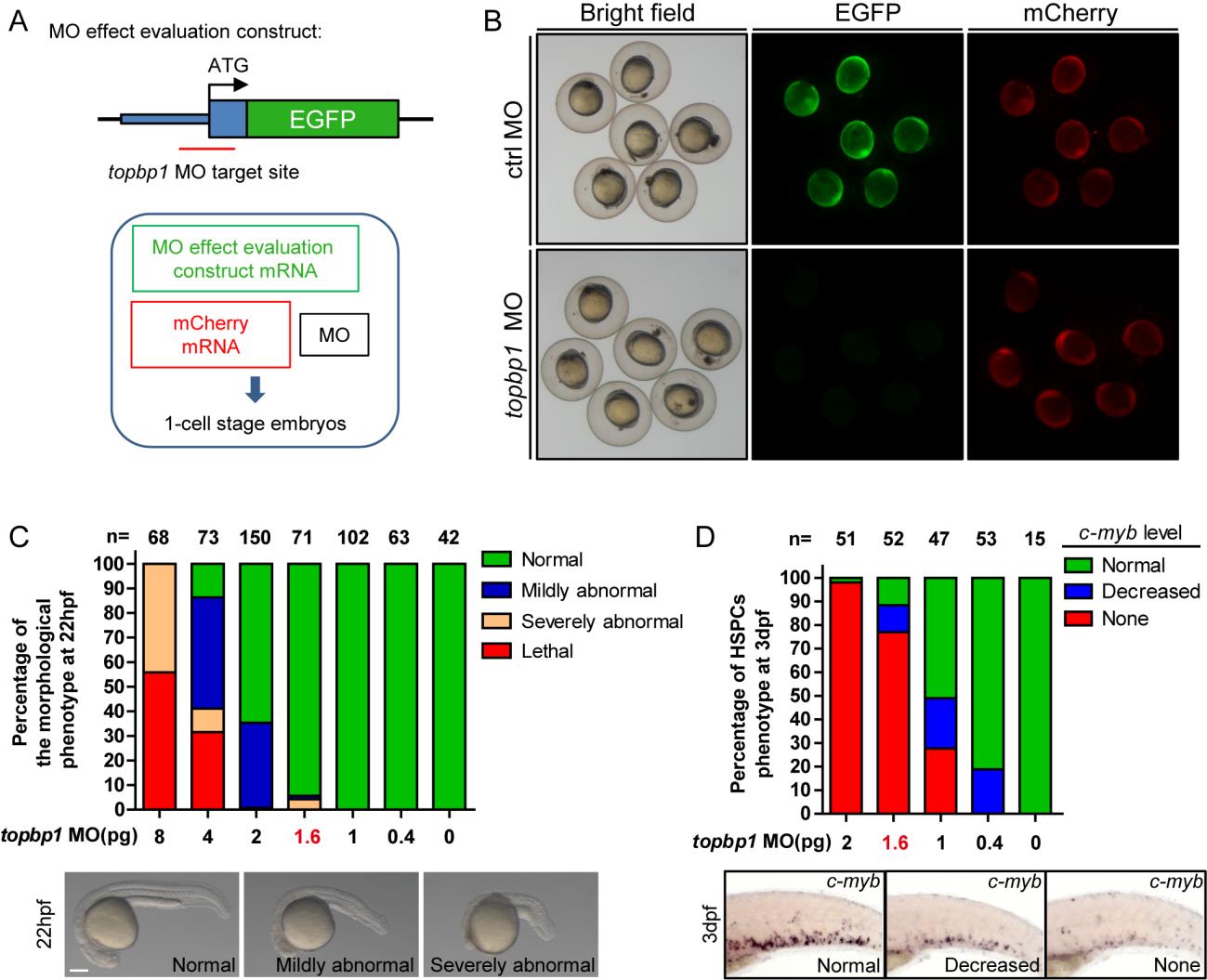
Fig. S3 The topbp1 morphants can phenocopy mutantcas003 embryos in a dose-dependent manner.
(A) Diagram of topbp1 MO knockdown effect evaluation construct. EGFP coding region was fused in frame to the 3′ end of a DNA fragment (blue boxes) containing topbp1 ATG MO targeting site (red line). This construct was in vitro transcripted, and then co-injected with mCherry mRNA (50pg) and topbp1 MO (1pg) or control MO (1pg) into 1-cell stage embryos. (B) Fluorescence of the 9hpf embryos in the topbp1 knockdown effect evaluation assay. topbp1 MO (upper), instead of control MO (down), can knockdown the expression of EGFP without affecting mCherry fluorescence. Left column, bright field; middle column, EGFP; right column, mCherry. (C) Quantitation of 22hpf morphology of the wild-type embryos injected with a gradient dose of topbp1 MO. Injection with more than 1.6pg topbp1 MO can induce abnormal morphogenesis. (D) Quantitation of the c-myb WISH analysis of embryos injected with a gradient dose of topbp1 MO at 3dpf. The topbp1 morphants can phenocopy topbp1cas003 mutants with 1.6–2 pg injection dosage without causing morphological defect.