
|
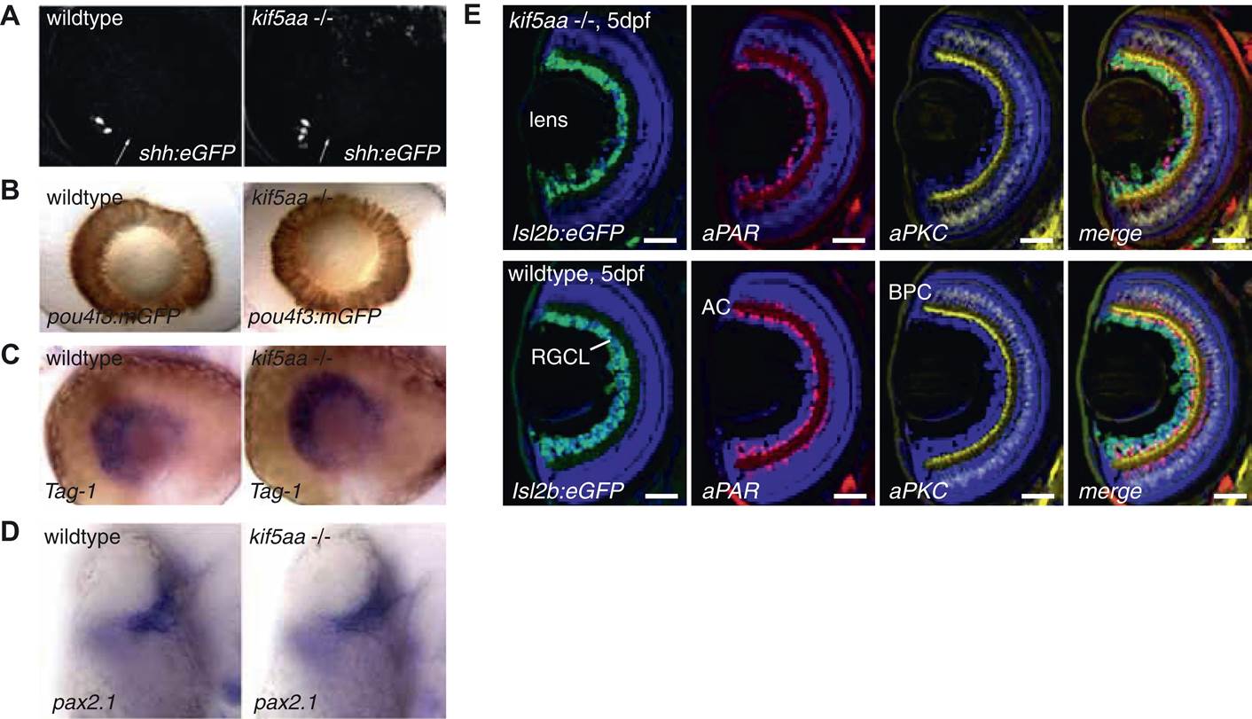
Fig. 2, S1 Patterning of the mutant retina and neurogenesis is not affected in mutants.
(A) The expression of the Tg(shh:eGFP) transgene marks the onset of neurogenesis (white arrow) in the developing retina (Shkumatava et al., 2004) and its expression is not altered in kif5aa mutant retinae. (B) RGC differentiation marked by Tg(pou4f3:mGFP) (Xiao et al., 2005) expression is not affected. (C) The glycosylphosphatidyl inositol (GPI)-anchored protein of the immunoglobulin (Ig) superfamily Tag-1 I is expressed in nasal RGCs (Lang et al., 2001). In situ staining shows that nasal patterning is identical in wild-type and kif5aa mutant retinae. (D) Pax2.1, whose expression is restricted to the optic stalk and retinal cells around the choroid fissure and that mediates optic stalk and chiasm formation in fish (Krauss et al., 1991; Puschel et al., 1992) shows a normal expression pattern in kif5aa mutants. (E) Cryosection of Tg(Isl2:Gal4, UAS:eGFP) transgenic wild-type and mutant retinae and immunostaining against eGFP (RGCs) (Ben Fredj et al., 2010), Parvalbumin (marking amacrine cells) (Godinho et al., 2005) and protein kinase C (marking bipolar cells) (Godinho et al., 2005) shows that all cell types are present in the right retinal layer. Blue = DAPI. Scale bar = 10 µm. RGCL = RGC layer, AC = amacrine cells, BPC = bipolar cells.