
|
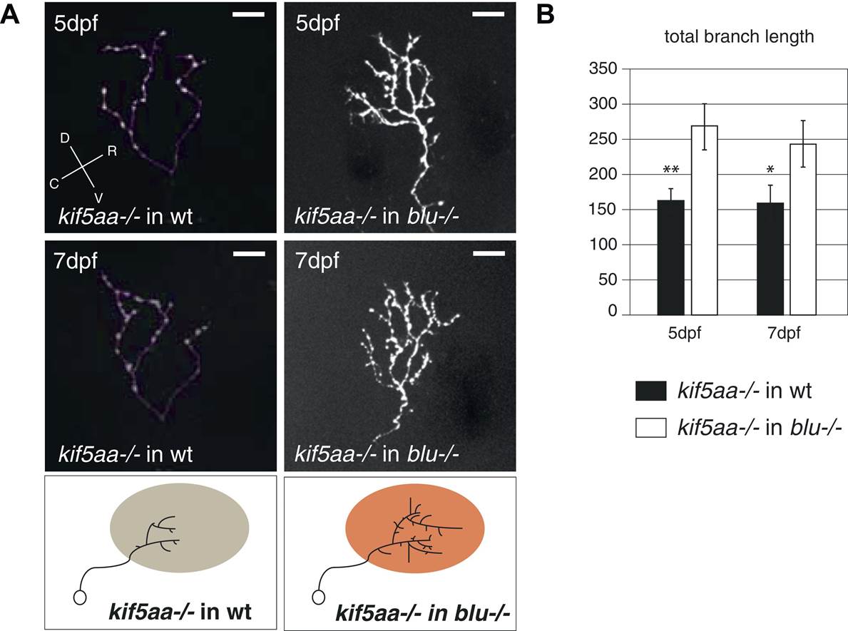
Fig. 8, S2 Transplantation of kif5aa mutant RGCs into a blumenkohl mutant acceptor leads to an increased growth compared to transplantation into a wild-type acceptor.
(A) Representative pictures of single in vivo imaged RGC axons after blastula stage transplantions from kif5aa mutant donors into a wild-type tectum (left panel) and from kif5aa mutants into a blumenkohl mutant tectum (right panel). The same cell was analyzed at 5 dpf (upper panel) and 7 dpf (middle panel). Scale bars = 20 µm. Schematics of RGC arbor complexity and size in the lower panel. In orange: Ntf3 overexpressing blumenkohl mutant tectum. D = dorsal, V = ventral, R = rostral, C = caudal. Pictures in the left panel are identical to Figure 8. (B) Quantification of total branch length of transplanted RGC axons at 5 and 7 dpf. The reduced size of kif5aa mutant axonal arbors when growing into a wild-type tectum is partially rescued when transplanted into a blumenkohl mutant environment (p < 0.05) (5 dpf: n = 35, 8; 7 dpf: n = 19, 8). The graph for kif5aa mutant cells into a wild-type host is identical to Figure 8.