
|
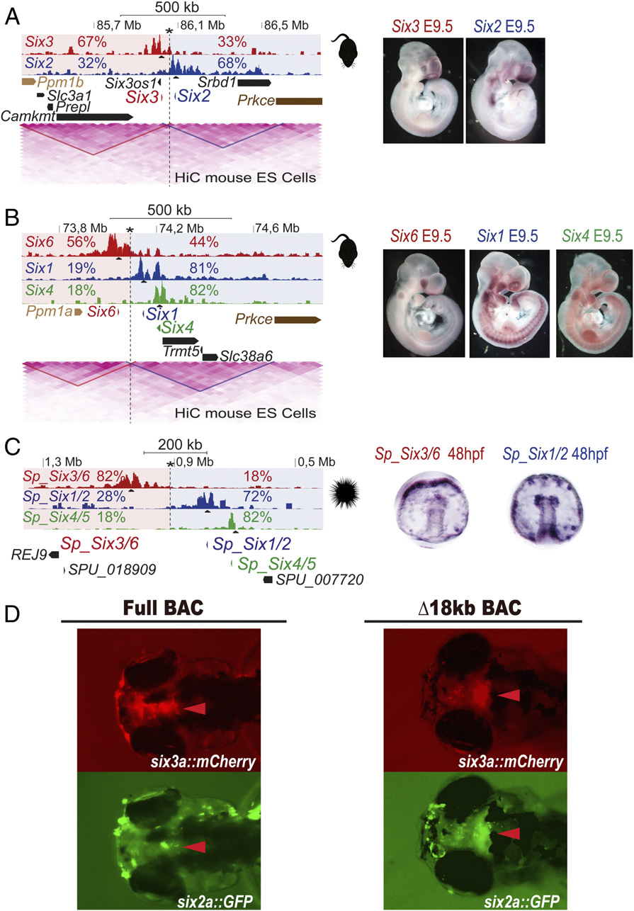
Fig. 3
Conserved subdivision of Six clusters in two 3D compartments along deuterostome evolution. (A–C) 4C-seq in whole mouse (A and B) or sea urchin (C) embryos from the different Six genes as viewpoints. Border regions are indicated by an asterisk, and the two 3D compartments are shaded in red and blue. Contact percentages for each gene on the two 3D compartments is shown. In murine clusters, HiC data from mouse ES cells is shown below. HiC data show that clusters are divided into the two TADs also defined by 4C-seq. Border regions identified by 4C-seq coincide in both clusters with TADs borders. (Right) Expression patterns of the different genes. (D) Transient transgenic fish embryos injected with the full six2a-GFP/six3a-mCherry BAC (Left) or a TAD-deleted version (Δ18kb) of the same BAC (Right) at 72 h hpf. Arrowheads point to the brain expression domain characteristic of the six3a gene.