
|
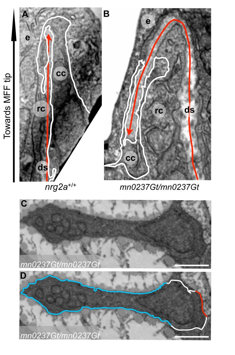
Fig. S4 Cleft cells in nrg2a mutants are largely unaffected, whereas ridge cells display expanded basal and reduced apical domains.
Transmission electron micrographs (TEM) of distal-most region of dorsal MFF of wild-type (A) and nrg2a mutant (mn0237Gt/mn0237Gt) (B-D) embryos at 36 hpf (A, B) or 52 hpf (C, D). (A, B) Cleft cell (cc) morphogenesis creates the cleft, an invagination of the nascent dermal space (ds, red arrow) into the cleft cell. White lines trace cleft cell boundaries; red arrow termini (red arrowheads) indicate termination of the dermal space within the cleft. The nrg2a mutant (B) has an intact cleft cell with normal morphology. (C, D) Representative example of a ridge bulging into the dermal space, consisting of a single ridge cell with an extended basal border (blue; D) and a noticeably reduced apical border (red, D). Lateral borders are in white (D). For clarity, identical images are shown side by side with (D) and without (C) marked ridge cell borders. Magnification: 10,000X, scale bar: 2 µm. (A-D) 36 hpf; (E-F) 2 dpf. Abbreviatiations: cc, cleft cell; ds, dermal space; e, EVL cell; rc, ridge cell.