
|
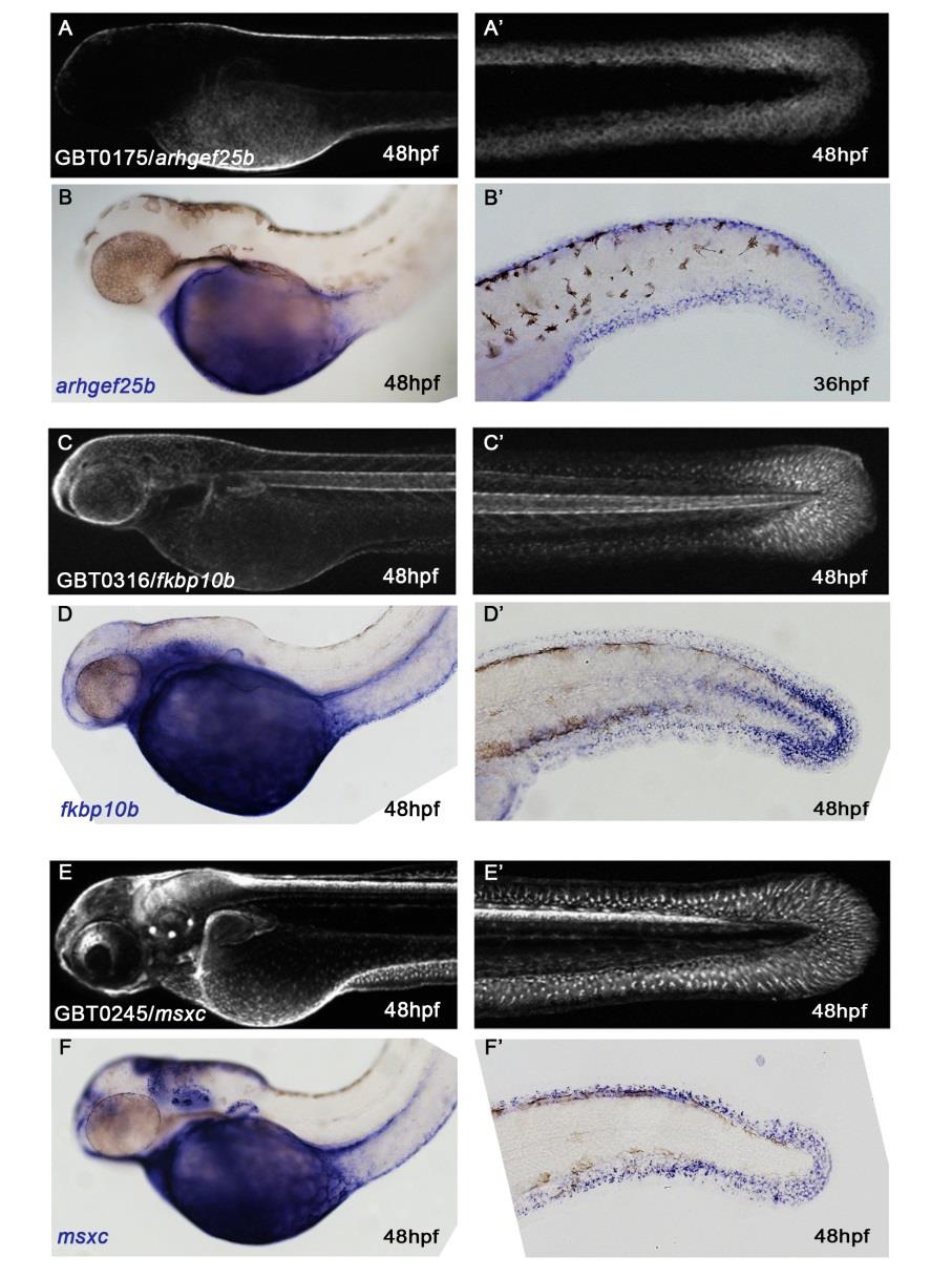
Fig. S1 mRFP fusion protein distribution in GBT lines recapitulates the endogenous expression patterns of arhgef25b, fkbp10b, and msxc.
Panels (A, A′, C, C′, E, E′) show the same in vivo fluorescence images of mRFP localization in GBT lines arhgef25bmn0175Gt (A, A′), fkbp10bmn0316Gt (C, C′) and msxcmn0245Gt (E, E′) as in Fig 2E, 2F, 2K, 2L, 2M, and 2N, respectively. Panels underneath show images of wild-type embryos of comparable stages (indicated in hpf) and orientations following whole-mount in situ hybridization (WISH) with arhgef25b (B, B′), fkbp10b (D, D′), or msxc (F, F′) RNA probes. mRFP localization in the epidermis covering the yolk in the arhgef25bmn0175Gt gene-break allele (A) corresponds to endogenous arhgef25b gene expression (B). Both arhgef25bmn0175Gt mRFP localization and endogenous arhgef25b gene expression are also observed in the MFF (A′, B′). Furthermore, just as the gene-break alleles fkbp10bmn0316Gt (C, C′) and msxcmn0245Gt (E, E′) show mRFP localization in fin mesenchymal cells (FMCs), fkbp10b and msxc genes show endogenous FMC expression (C-F′; also compare with MsxCmn0245Gt-mRFP fusion protein localization in Fig 3E–3H). mRFP localization to the pectoral fin buds observed in the fkbp10bmn0316Gt allele (C) parallels endogenous fkbp10b expression in the pectoral fin buds (D). fkbp10b also shows expression in the posterior region of the notochord (D′). The weaker WISH signal in anterior (earlier specified) regions of the notochord compared to the mRFP signal (C′) points to transient expression of the endogenous gene in notochord cells, and higher stability of the GBT-generated mRFP fusion protein than the endogenous transcript. The endogenous msxc gene is also expressed in the maculae of the inner ear and in the pectoral fin buds (F), confirming that the msxcmn0245Gt GBT allele and its resulting mRFP fusion protein also recapitulate endogenous expression in tissues other than the skin (E).