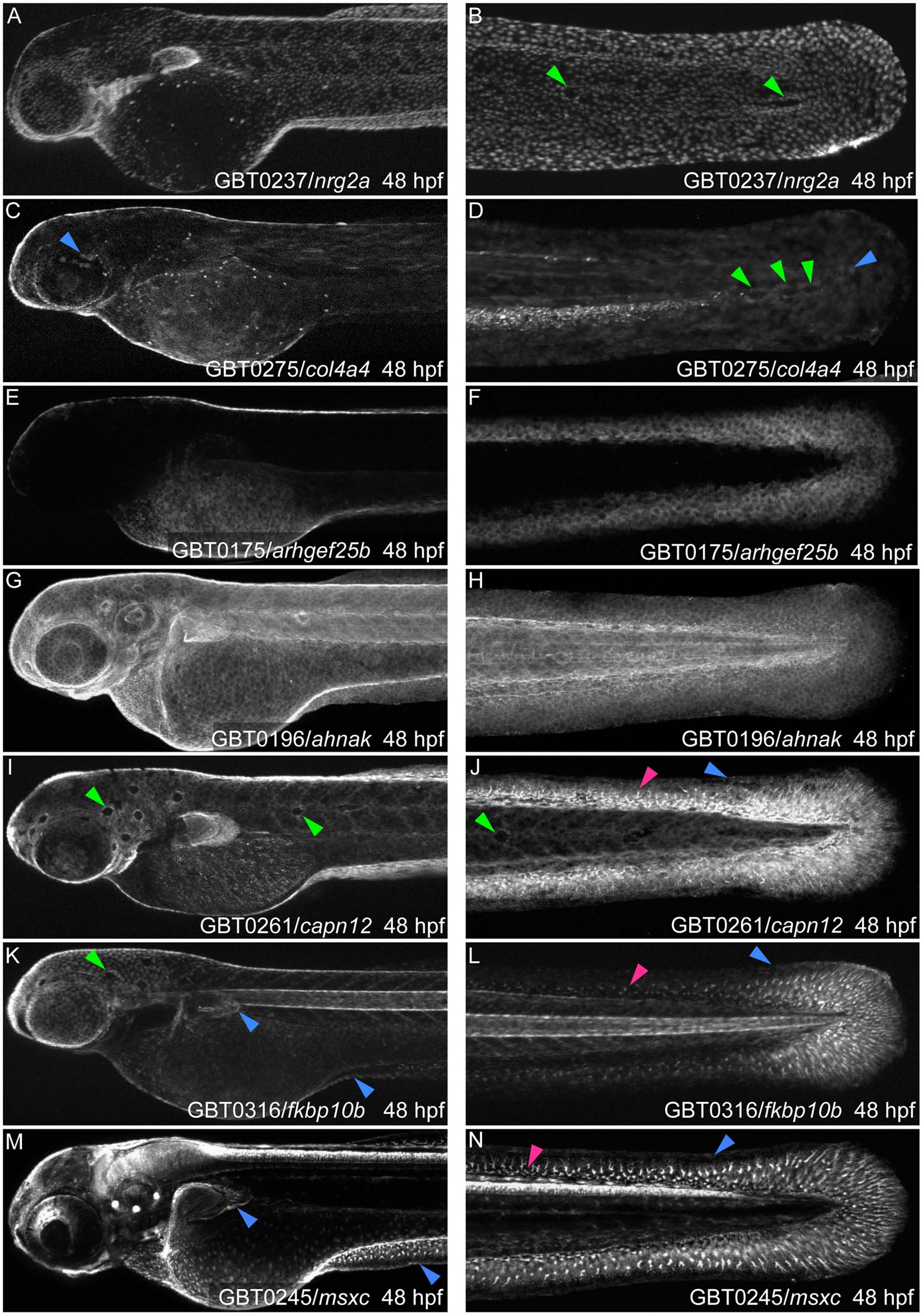
Fig. 2 GBT protein trapping identifies integument loci with epidermal or fin mesenchymal expression.
As identified by mRFP localization, seven GBT alleles have unique epidermal expression patterns emphasizing epidermal continuity over the body and larval fin folds. They are: (A, B) nrg2amn0237Gt, (C, D) col4a4mn0275Gt, (E, F) arhgef25bmn0175Gt, (G, H) ahnakmn0196Gt, (I, J) capn12mn0261Gt, (K, L) fkbp10bmn0316Gt, and (M, N) msxcmn0245Gt. (B, D, I, J, K) Four lines, nrg2amn0237Gt, col4a4mn0275Gt, capn12mn0261Gt, and fkbp10bmn0316Gt, have “holes” or “gaps” in their epidermal pattern (green arrowheads) where neuromasts embedded in the basal layer exclude the mRFP-positive basal keratinocytes. (A, G, I, K, M) nrg2amn0237Gt (A), ahnakmn0196Gt (G), capn12mn0261Gt (I), fkbp10bmn0316Gt (K), and msxcmn0245Gt (M) are also expressed in the pectoral fin folds. (C-D, J, L, M-N) col4a4mn0275Gt, capn12mn0261Gt, fkbp10bmn0316Gt, and msxcmn0245Gt epidermal expression (blue arrowheads) can be difficult to discern among other expression pattern components. (I-N) capn12mn0261Gt, megf6amn0316Gt, and msxcmn0245Gt also show expression in fin mesenchymal cells (pink arrowheads). (C, D, K, L, M, N) Several lines are also expressed in other tissues. (C, D) col4a4mn0275Gt appears in myotomes and the vascular plexus. (K, L) fkbp10bmn0316Gt is seen in the notochord. (M, N) msxcmn0245Gt is strongly expressed in the brain, spinal cord, and sensory maculae. hpf, hours post-fertilization. Comparisons of the mRFP localization patterns with the expression pattern of the endogenous genes are shown in Supporting Information (S1 Fig)

