
Fig. 1
- ID
- ZDB-IMAGE-150810-1
- Publication
- Evason et al., 2015 - Identification of Chemical Inhibitors of β-Catenin-Driven Liver Tumorigenesis in Zebrafish
- All Figures
- Figures for Evason et al., 2015
|
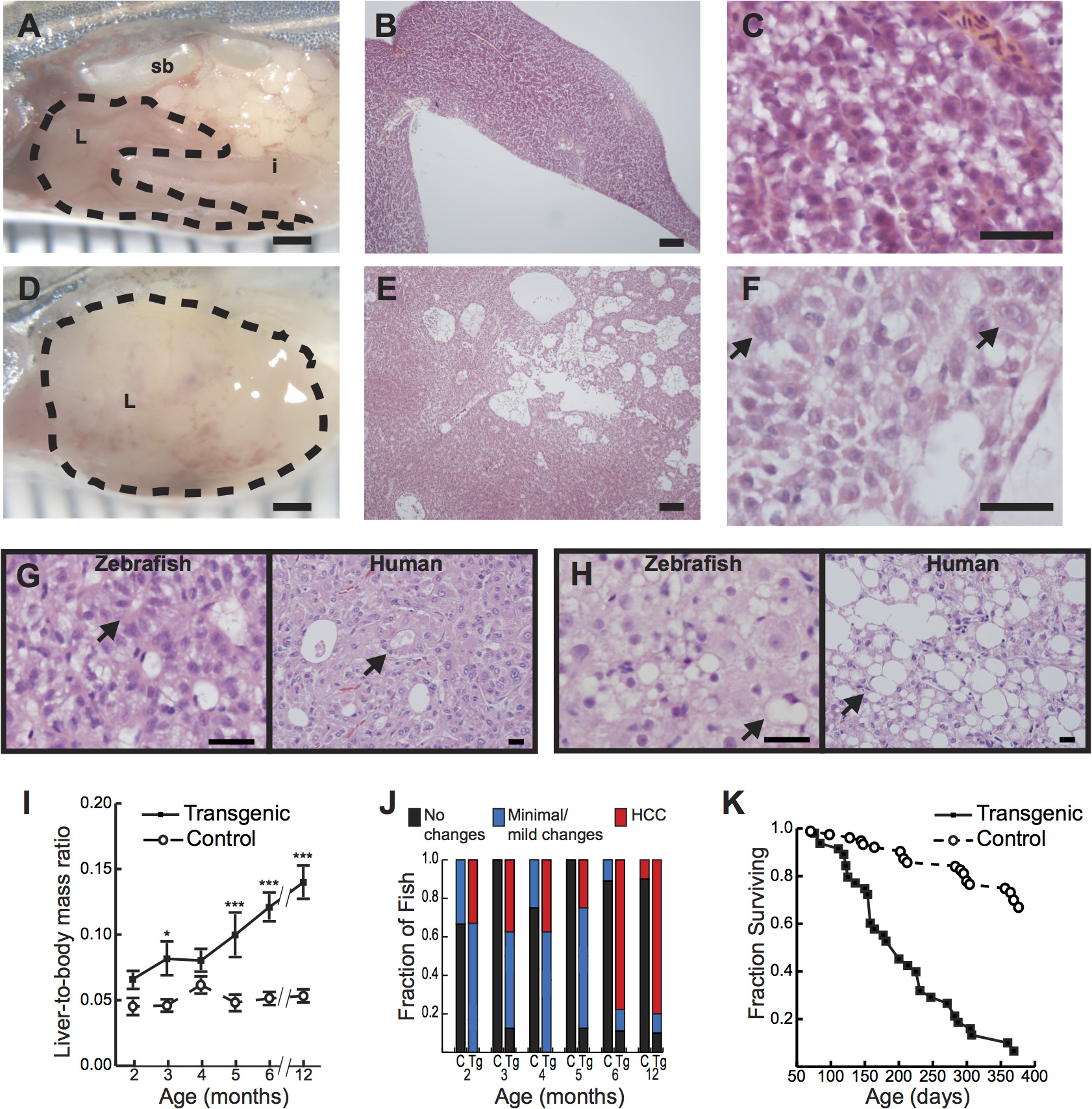
Fig. 1 Hepatocyte-specific expression of activated β-catenin results in liver enlargement, hepatocellular carcinoma (HCC), and decreased survival in adult zebrafish.
(A-C) Control 4-month-old zebrafish showing the liver (L, outlined) positioned in the body cavity near the intestine (i) and swim bladder (sb)(A). Sections show an orderly arrangement of hepatocytes (B-C). (D-F) Transgenic 4-month-old zebrafish showing an enlarged liver (D) with disorganized architecture (E) and atypical cells (arrows, F). (G-H) Transgenic 6-month-old (G) or 4-month-old (H) zebrafish and human HCC showing architectural disruption with scattered pseudoglands (arrows, G) and evidence of intracellular lipid accumulation (arrows, H). Hematoxylin and eosin stains; scale bars, 1 mm (A, D), 100 µm (B, E), 25 µm (C, F), and 20 µm (G, H). (I) Graph showing average liver size normalized to total body mass, ± standard error of the mean (SEM). Asterisks indicate p-values for ANOVA comparing transgenic zebrafish (N = 56) to control siblings (N = 51) at the same time point: *, p<0.05; ***, p<0.001. (J) Livers of transgenic zebrafish (Tg, N = 49) and control siblings (C, N = 37) were examined microscopically, and architectural and cytological changes were scored. HCC was significantly more common in transgenic zebrafish than in controls (p<0.001, Fisher’s exact test). (K) Kaplan-Meier survival curves comparing adult survival of transgenic zebrafish (N = 51) and control siblings (N = 85); p<0.001, logrank test.