
|
Fig. S8
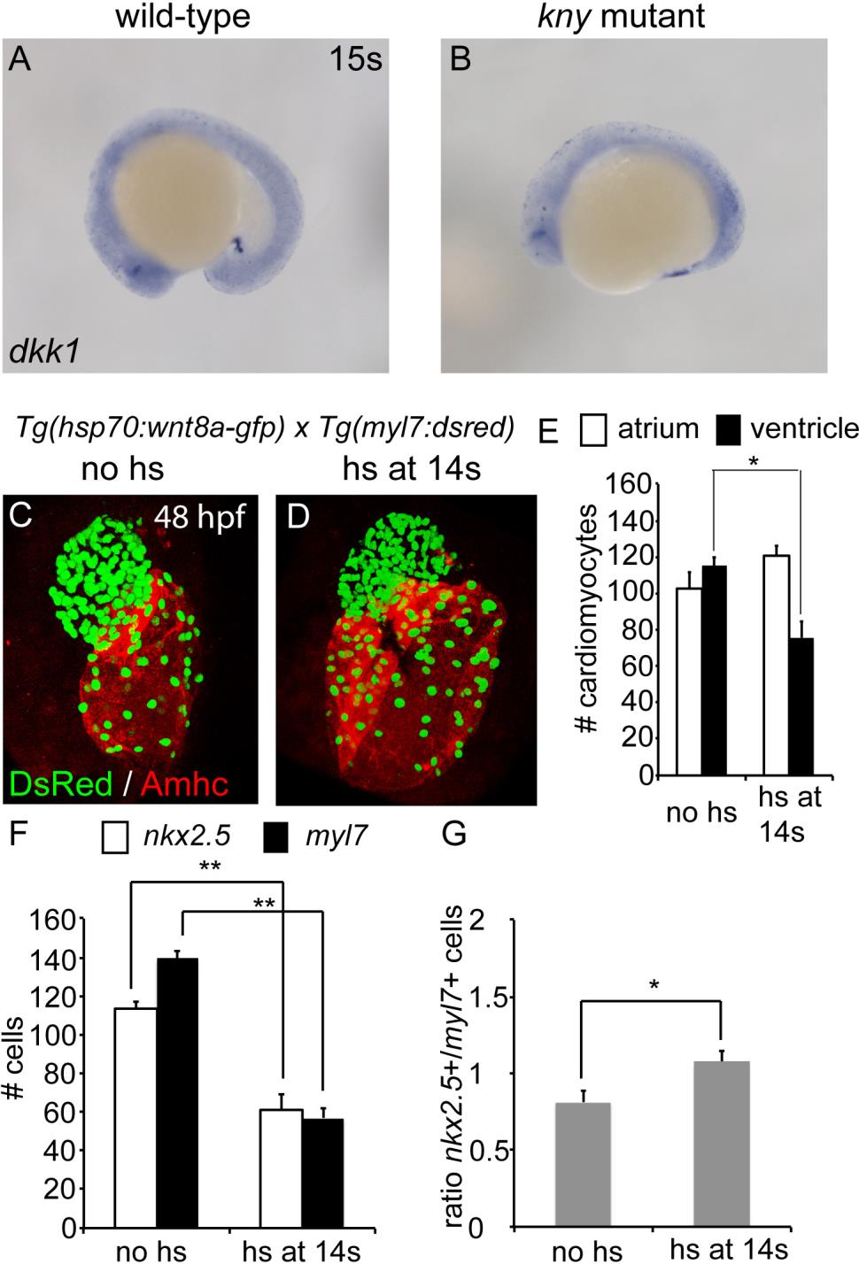
Related to Figure 4: Elevation of canonical Wnt signaling after cardiac specification leads to a reduction of ventricular cardiomyocytes. (A,B) Lateral view of wild-type siblings and kny/gpc4 mutants after in situ hybridization for dkk1 at the 15 somite stage. (C,D) Hearts at 48 hpf of embryos derived from the Tg(hsp70:wnt8a-gfp) line crossed to Tg(myl7:dsred), stained for DsRed (false colored in green) and Amhc (red). (E) Cardiomyocyte numbers of corresponding hearts (n=3). (F) Quantification of nkx2.5+ and myl7+ cells in the ventricles of embryos after wnt8a induction (n=3). (G) Ratio of nkx2.5+/myl7+ cells in corresponding hearts after Wnt8 induction. Results are represented as mean ±s.e.m. Asterisks represents statistical significance according to a paired t-test: * P<0.05; ** P<0.01.