
|
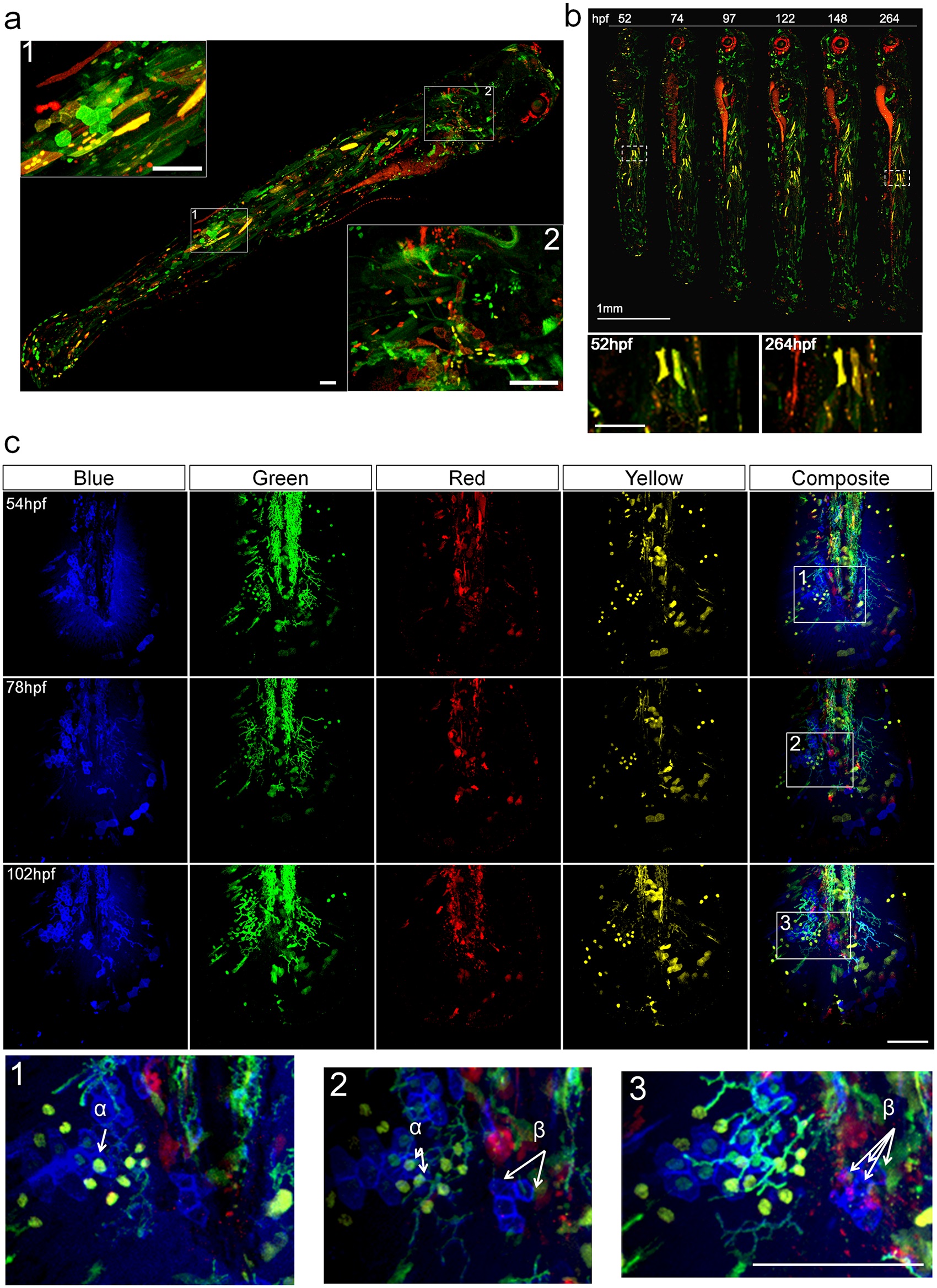
Fig. 2
Spatial temporal coverage and stability of Multibow labeling.
a. Spatial and cell type coverage of Multibow. The embryo was injected with 6 Multibow colors (mR/mG/nR/nG/R/G) at single cell stage and heat-shocked at 1 day-post-fertilization (dpf) for 2 hours. The whole 4dpf larva was imaged in 2 channels (G/R). Positive cells can be seen distributed from head to tail throughout the larva, indicating high spatial coverage. In inserts 1 and 2, distinctly shaped skin, muscle, mesenchymal and neural cells can be observed by cytoplasmic or membrane Multibow labeling. Scale bars: 100µm. b. Temporal stability of labeling. The embryo was injected with 6 Multibow colors (mR/mG/nR/nG/R/G) at single cell stage and heat-shocked at 1 day-post-fertilization (dpf) for 2 hours. The same embryo was imaged once per day to 11dpf. The persistence of labeling indicates genomic insertion of Multibow cassettes. Red patches around the eye and along the gut are auto-fluorescence. Enlarged views of white boxed areas show that the area is stably fluorescent. Scale bar in enlarged views: 100µm. c. Label stability of color codes over time. The embryo was injected with 12 (B/G/Y/R) Multibow constructs at one cell stage. Heat-shock of this tg(hsp70:cerulean-cre) individual was at 30hpf (duration: 2 hours). Its developing larval tail fin was imaged every 24 hours starting at 54hpf using four channels (B/G/Y/R). The color codes of the cells remain unchanged despite fluorescent intensity differences at different days, allowing identification of the same cells/clones(e.g., α and β, shown in enlarged regions marked by white boxes). Color codes: α: nG/nY; β: mB. Scale bar: 100µm. See also Fig d in S2 Fig.