
IMAGE
Fig. 7, S2
- ID
- ZDB-IMAGE-150713-34
- Genes
- Antibodies
- Publication
- Fadeev et al., 2015 - Tight junction protein 1a regulates pigment cell organisation during zebrafish colour patterning
- All Figures
- Figures for Fadeev et al., 2015
Image
|
Figure Caption
Fig. 7, S2
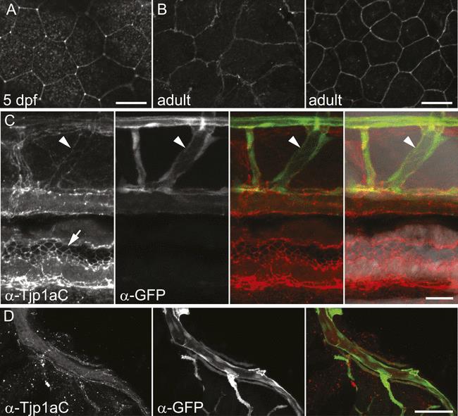
(A) Signal in epithelium of wild type 5 dpf larva stained with α-Tjp1aC. Scale bar: 20 µm. (B) Signal in two layers of adult wild type epithelium (about 7 µm apart) stained with α-Tjp1aC st. Scale bar: 20 µm. (C) Double staining of whole-mount Tg(kdrl:GFP) 5 dpf larvae with α-Tjp1aC and α-GFP demonstrates the expression of Tjp1a in blood vessels (arrowheads) and intestinal epithelium (arrows). Scale bar: 20 µm. (D) α-Tjp1aC staining shows expression of Tjp1a in vasculature of adult Tg(kdrl:GFP) animal. Scale bar: 50 µm.
Figure Data
Acknowledgments
This image is the copyrighted work of the attributed author or publisher, and
ZFIN has permission only to display this image to its users.
Additional permissions should be obtained from the applicable author or publisher of the image.
Full text @ Elife