
Fig. S5
- ID
- ZDB-IMAGE-150708-30
- Publication
- Harrison et al., 2015 - Chemokine-guided angiogenesis directs coronary vasculature formation in zebrafish
- All Figures
- Figures for Harrison et al., 2015
|
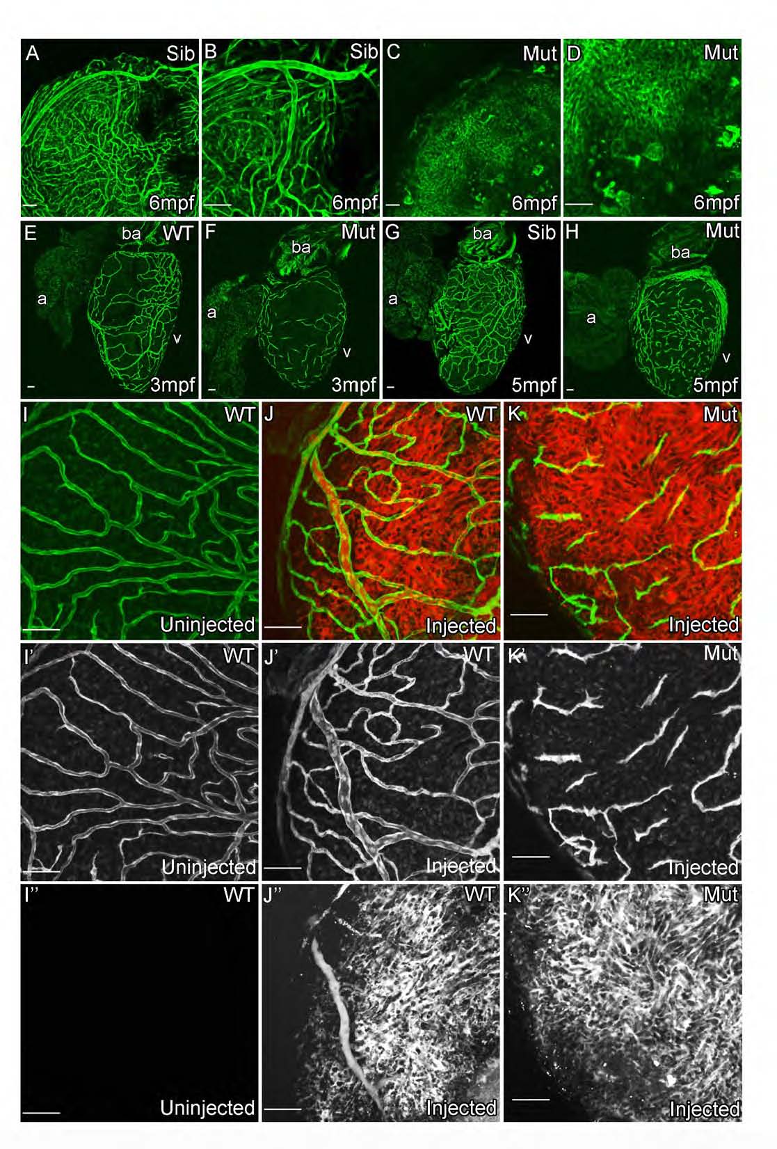
Fig. S5 cxcr4a mutant zebrafish fail to develop functional coronary vasculature. (Related to Figure 5)
kdrl:grcfp allows visualization of the endocardium, bright atrial vessels and weaker expressing non-arterial vessels in 6mpf sibling adult zebrafish (A and B). This network of vessels is not detectable in the cxcr4a mutant zebrafish; instead unorganized endothelial cells are detectable on the surface of the heart (C and D). These cells fail to form vessels and remain as isolated cells into adulthood (E-H). Angiographs suggest that there is no major supply of blood to the surface of the heart in the mutant other than the systemic flow [I-K; Green, fli1a:EGFP (I′-K′); Red, retro-orbitally injected Rhodamine-Dextran (I′′-K′′)]. Scale bars, 50 µm.
Reprinted from Developmental Cell, 33, Harrison, M.R., Bussmann, J., Huang, Y., Zhao, L., Osorio, A., Burns, C.G., Burns, C.E., Sucov, H.M., Siekmann, A.F., Lien, C.L., Chemokine-guided angiogenesis directs coronary vasculature formation in zebrafish, 442-54, Copyright (2015) with permission from Elsevier. Full text @ Dev. Cell