
Fig. S4
- ID
- ZDB-IMAGE-150708-29
- Publication
- Harrison et al., 2015 - Chemokine-guided angiogenesis directs coronary vasculature formation in zebrafish
- All Figures
- Figures for Harrison et al., 2015
|
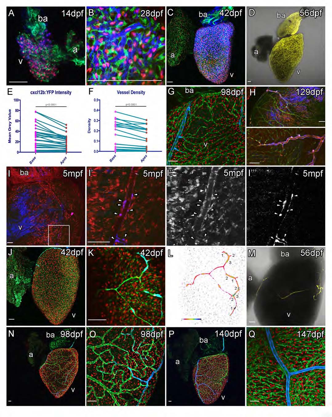
Fig. S4 cxcl12b:YFP and cxcr4a:YFP expression during vessel formation. (Related to Figure 4)
Prior to the emergence of vessels, cxcl12b:YFP (pseudo colored blue in A, B, C, G, H, I; yellow in D) is detected in ventricular cardiomyocytes that also express myl7:DsRed2-NLS and are flanked by fli1a:EGFP positive endocardial cells (AC). Expression of cxcl12b:YFP is stronger in the base than the apex (E, between 14dpf and 84dpf, n=36, measurements paired within hearts) and coronary vasculature is significantly more dense in the base of the ventricle (F, between 7 and 420dpf, n=153, means of 22 time points are paired), both p<0.0001 (Wilcoxon matched-pairs signed rank test). After the formation of coronary vessels expression is observed in mural cells (G-I). tcf21:CreERt2, ubb:LoxPEGFP- LoxP-mCherry, cxcl12b:YFP embryos are treated with 4OHT to label the embryonic endocardium. mCherry-positive, epicardium (tcf21+) derived cells line the vessels and are cxcl12b:YFP positive (I, box enlarged to I′, I′′ mCherry, I′′′ YFP). Endothelial cells that migrate out over the ventricular cardiomyocytes express cxcr4a:YFP (J, K, L and M). cxcr4a:YFP expression becomes down regulated as vessels form (N-Q). cxcr4a:YFP expression is maintained in large vessels and this expression persists late into adulthood after the coronary vasculature has formed (P and Q). cxcr4a:YFP (peudocolored blue), myl7:DsRed2-NLS (red) and fli1a:EGFP (green) in J, K, N-Q. Colorimetric display is used to show cxcr4a:YFP intensity (L). Single cxcr4a:YFP transgenic fish (M; YFP, yellow; PMT, grey). Scale bars, 50 µm.
Reprinted from Developmental Cell, 33, Harrison, M.R., Bussmann, J., Huang, Y., Zhao, L., Osorio, A., Burns, C.G., Burns, C.E., Sucov, H.M., Siekmann, A.F., Lien, C.L., Chemokine-guided angiogenesis directs coronary vasculature formation in zebrafish, 442-54, Copyright (2015) with permission from Elsevier. Full text @ Dev. Cell