
Fig. S1
- ID
- ZDB-IMAGE-150708-26
- Publication
- Harrison et al., 2015 - Chemokine-guided angiogenesis directs coronary vasculature formation in zebrafish
- All Figures
- Figures for Harrison et al., 2015
|
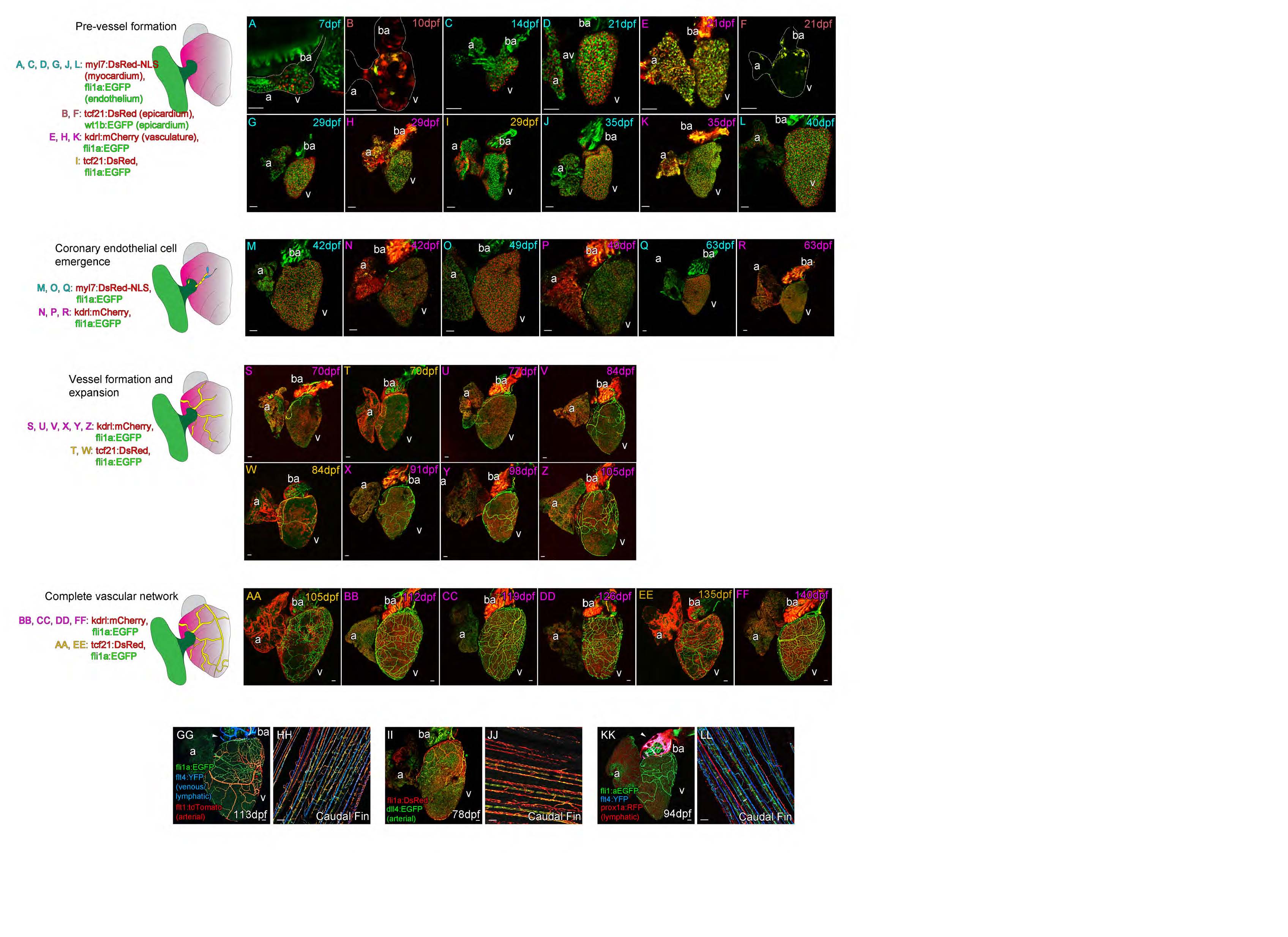
Fig. S1 Postembryonic zebrafish coronary vessel development. (Related to Figure 1)
Four different transgenic lines were used in different combinations to characterize the development of coronary vessels in zebrafish from 7dpf to 140dpf; fli1a:EGFP; myl7:DsRed2-NLS (labeled blue: A, C, D, G, J, L, M, O and Q), wt1b:EGFP; tcf21:DsRed (labeled maroon: B and F), fli1a:EGFP; kdrl:mCherry (labeled pink: E, H, K, N, P, R, S, U, V, X, Y, Z, BB, CC, DD, FF), fli1a:EGFP; tcf21:DsRed (labeled orange: I, T, W, AA, EE).
Pre-vessel formation (7 to 40dpf). After hatching the heart is composed of three layers, the fli1a:EGFP endocardium (A), cmlc:nRFP positive myocardium (A) and the epicardium partially labeled with wt1b:EGFP and tcf21:DsRed (B). The heart increases in size dramatically through the first six weeks of life (A-L) during which time the myocardium expands and the tcf21-poisitive epicardium partially covers the surface of both the atrium (a) and ventricle (v) (B, F, I). The bulbus arteriosus (ba) strongly expresses kdrl:mCherry as does the endocardium (E, H, K), which is visible through the myocardium. There are no endothelial cells (fli1a:EGFP) on the surface of the myocardium (cmlc:nRFP, A, C, D, G, J and L).
Coronary endothelial cell emergence (42 to 63dpf). Following this expansion in the six weeks after fertilization, endothelial cells are found on the surface of the heart and the coronary vasculature begins to form (M-R). These endothelial cells are found on the outside of the myocardium (cmlc:nRFP, M, O, Q) and express kdrl:mCherry (N, P, R). They are first observed at the juncture of the atrium and ventricle (M-O) and then connected from this point over the base of the ventricle (P-R).
Vessel formation and expansion (70 to 105dpf). The heart continues to grow into adulthood as it does so the vasculature continues to form over the surface of the ventricle, but not the atrium (S-Z). Endothelial cells remain connected to existing sprouts or vessels during this process (S-Z) and the heart becomes increasingly covered by tcf21-possitive epicardial cells as it proceeds (T and W).
Complete vascular network (105 to 140dpf). By 4 months post-fertilization the intricate network of vessels is formed, the pattern and density of vessels varies between adult zebrafish, but vessels are only observed on the ventricle of the heart (AA-FF).
A subset of endothelial cells of the coronary vasculature network express arterial markers flt1:tdtomato (GG) and dll4:EGFP (II), but none of the vasculature endothelial cells express venous marker flt4:YFP (GG and KK) nor lymphatic marker prox1:RFP (KK) at this stage. Expression of both prox1a:RFP and flt4:YFP is restricted to endothelium partially covering the BA (GG and KK, arrowheads). All four markers were found to be expressed in the adult fin of the same fish (flt1:tdtomato and flt4:YFP, HH; dll4:EGFP, JJ; prox1a:RFP and flt4:YFP, LL). Asterisk denotes artery. Scale bars, 50 µm.
Reprinted from Developmental Cell, 33, Harrison, M.R., Bussmann, J., Huang, Y., Zhao, L., Osorio, A., Burns, C.G., Burns, C.E., Sucov, H.M., Siekmann, A.F., Lien, C.L., Chemokine-guided angiogenesis directs coronary vasculature formation in zebrafish, 442-54, Copyright (2015) with permission from Elsevier. Full text @ Dev. Cell