
Fig. 7
- ID
- ZDB-IMAGE-150708-25
- Publication
- Harrison et al., 2015 - Chemokine-guided angiogenesis directs coronary vasculature formation in zebrafish
- All Figures
- Figures for Harrison et al., 2015
|
Fig. 7
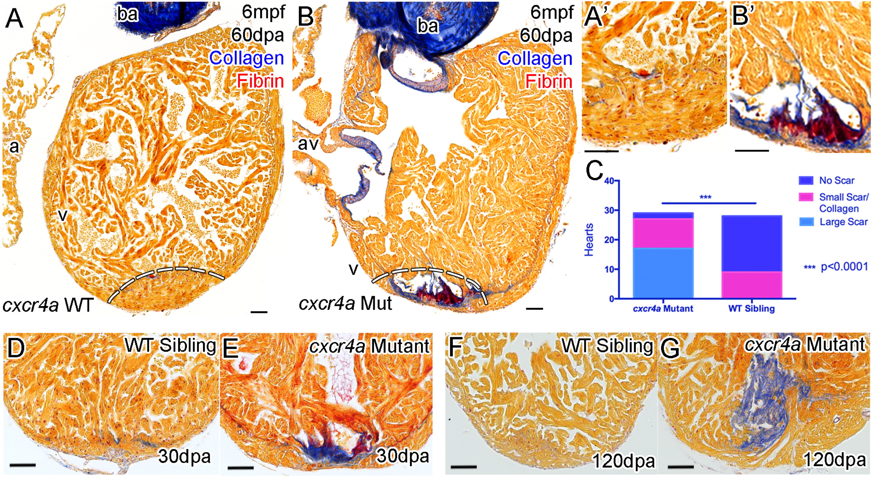
cxcr4a Mutant Hearts Fail to Regenerate following Resection of the Ventricle Apex
Zebrafish with functional Cxcr4a-Cxcl12b signaling and normal coronary vessel development completely regenerate resected ventricle tissue after 60 days (A, dashed line represents amputation plane, and A′). Adult cxcr4a mutants with disrupted coronary vessel development fail to regenerate the resected tissue resulting in the deposition and persistence of collagen (blue) and fibrin (red) (B, B′, E, and G). Quantification of scar formation at 60 dpa (C). cxcr4 mutant zebrafish have significantly more collagen deposition and scar formation compared to the siblings (p < 0.0001, chi-square). Scar formation is observed by 30 dpa (D and E) and persists beyond 120 dpa (F and G) with no apparent effect on survival. Scale bars represent 50 µm.
Reprinted from Developmental Cell, 33, Harrison, M.R., Bussmann, J., Huang, Y., Zhao, L., Osorio, A., Burns, C.G., Burns, C.E., Sucov, H.M., Siekmann, A.F., Lien, C.L., Chemokine-guided angiogenesis directs coronary vasculature formation in zebrafish, 442-54, Copyright (2015) with permission from Elsevier. Full text @ Dev. Cell