
Fig. 6
- ID
- ZDB-IMAGE-150708-24
- Publication
- Harrison et al., 2015 - Chemokine-guided angiogenesis directs coronary vasculature formation in zebrafish
- All Figures
- Figures for Harrison et al., 2015
|
Fig. 6
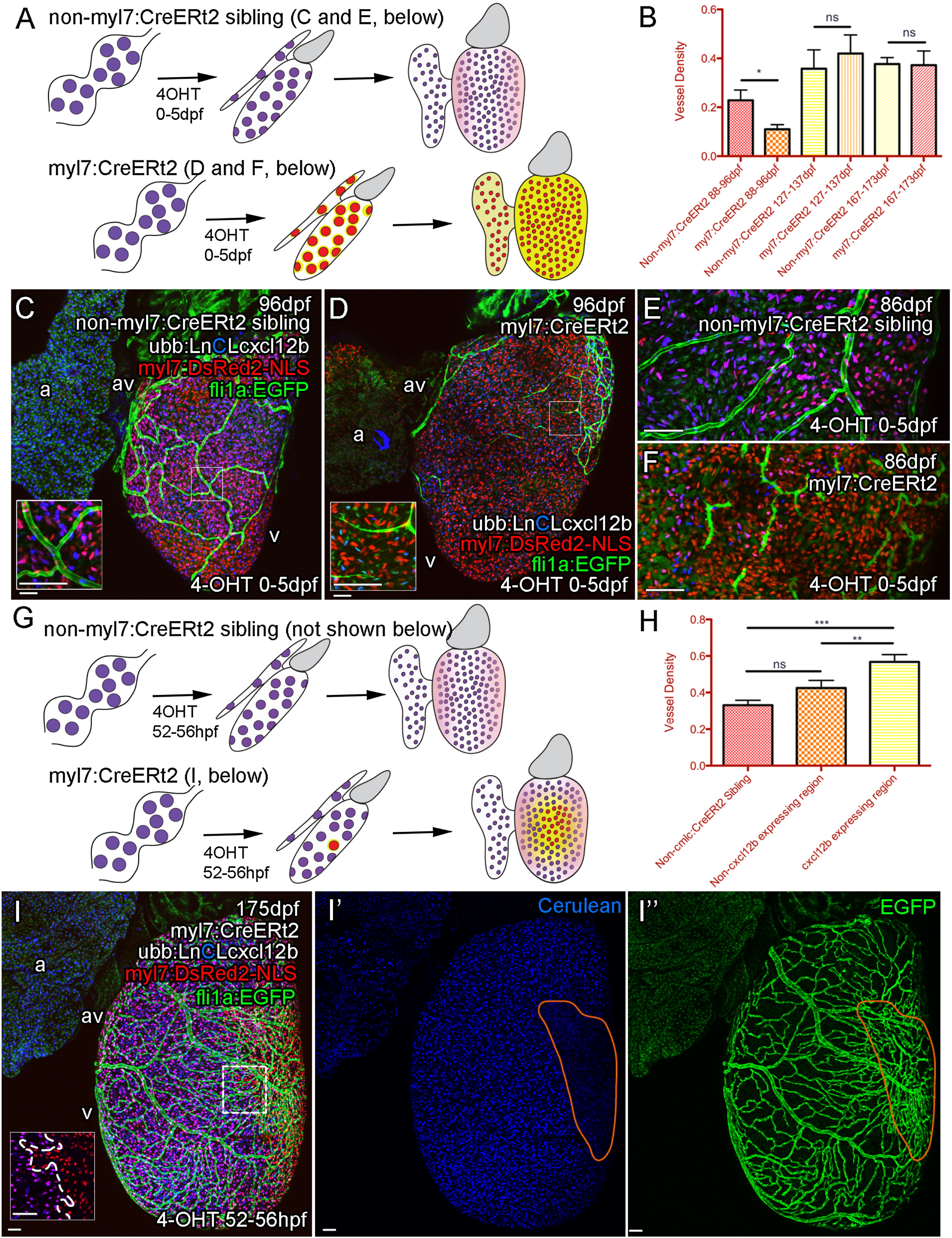
Cxcl12b Likely Provides Positional Information for Coronary Endothelial Cells
(A–E) Uniform overexpression of Cxcl12b in quadruple transgenic fish Tg(Ubb:LoxPH2B-Cerulean-LoxP-cxcl12b(ubb:LnCLcxcl12b); myl7:DsRed2-NLS; fli1a:EGFP; myl7:CreERt2). Schematic illustration of uniform overexpression of Cxcl12b. Prolonged treatment with 4OHT between 0 and 5 dpf resulted in efficient removal of the floxed H2B-Cerulean (blue) and expression of cxcl12b (yellow) in myl7-positive cardiomyocytes (red) such that any endogenous Cxcl12b signaling pattern may be obscured (pink) (A). Such overexpression of cxcl12b in cardiomyocytes also resulted in an initial disruption of vessel formation (B, quantification of vessel density, p < 0.05 t test, ± SEM; C, non-myl7:CreERt2 sibling control; D, cxcl12b overexpression) with endothelial cells becoming elongated and appearing spiculated (C and D, inset, E [control], and F [overexpression]). (G–I) Clonal overexpression of Cxcl12b in quadruple transgenic fish Tg(Ubb:LoxPH3B-Cerulean-LoxP-cxcl12b (ubb:LnCLcxcl12b); myl7:DsRed2-NLS; fli1a:EGFP; myl7:CreERt2). A 4-hr pulse of 4OHT results in partial activation of the myl7:CreERt2 transgene prior to cardiomyocyte clonal expansion. cxcl12b is then expressed from a localized source in adult zebrafish (G, I; inset from box; boundary between exogenous-cxcl12b expressing [DsRed-NLS positive] and non-expressing [H2B-Cerulean and DsRed-NLS double positive] cardiomyocytes is marked with a dashed line). There is an increase in density of vessels over and proximal to the region of cxcl12b expressing cardiomyocytes (H, I, I′, and I′′, region of cxcl12b-expressing (Cerulean-negative) cardiomyocytes demarcated by orange). (H) Quantification of vessel density. Representative image of clonal overexpression (I, I′, and I′′) with region of cxcl12b-expressing (Cerulean-negative) cardiomyocytes demarcated by orange. p < 0.01; p < 0.0001 t test. Scale bars represent 50 µm.
Reprinted from Developmental Cell, 33, Harrison, M.R., Bussmann, J., Huang, Y., Zhao, L., Osorio, A., Burns, C.G., Burns, C.E., Sucov, H.M., Siekmann, A.F., Lien, C.L., Chemokine-guided angiogenesis directs coronary vasculature formation in zebrafish, 442-54, Copyright (2015) with permission from Elsevier. Full text @ Dev. Cell