
Fig. 4
- ID
- ZDB-IMAGE-150708-22
- Publication
- Harrison et al., 2015 - Chemokine-guided angiogenesis directs coronary vasculature formation in zebrafish
- All Figures
- Figures for Harrison et al., 2015
|
Fig. 4
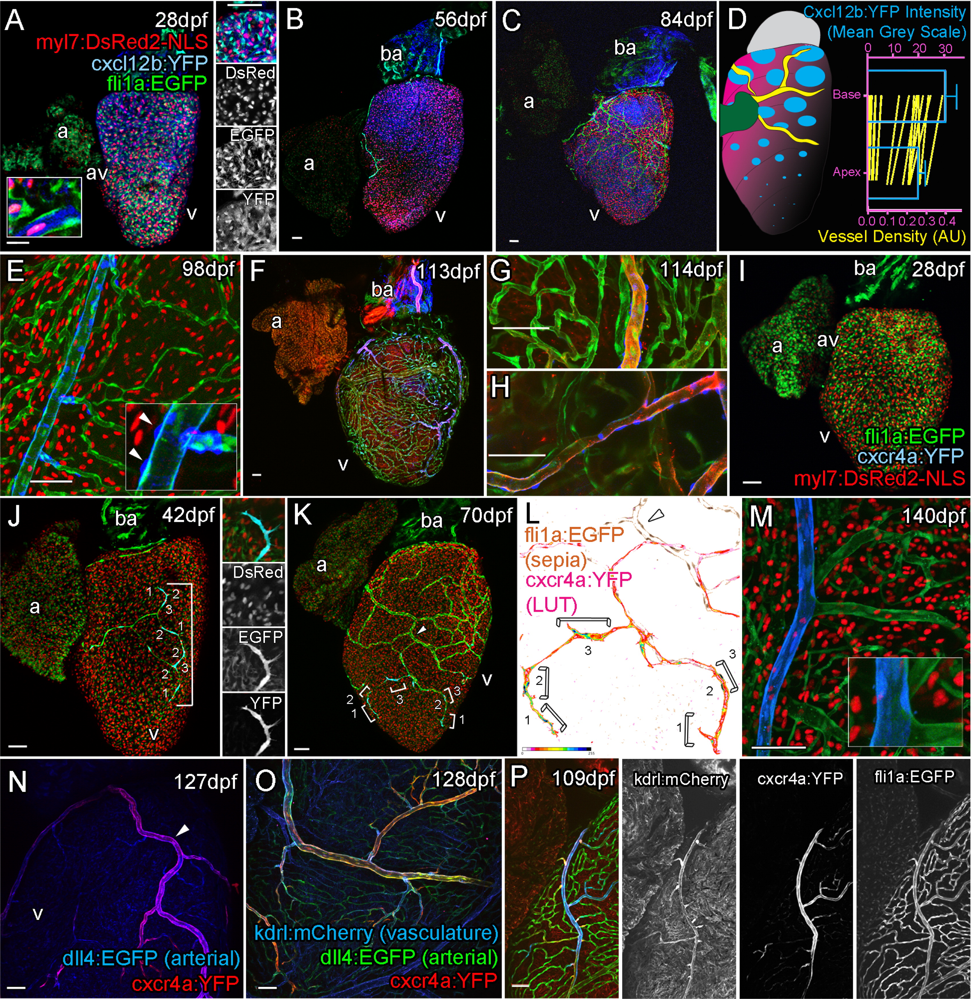
Cxcr4a-Cxcl12b Expression during Coronary Vessel Formation
Confocal images of transgenic fish Tg(myl7:DsRed2-NLS; fli1:EGFP; cxcl12b:YFP) (A–C, E), Tg(kdrl:mCherry; fli1:EGFP; cxcl12b:YFP) (F–H), Tg(myl7:DsRed2-NLS; fli1:EGFP; cxcr4a:YFP) (I–M), Tg(dll4:EGFP; cxcr4a:YFP) (N), Tg(dll4:EGFP; cxcr4a:YFP; kdrl:mCherry) (O), and Tg(kdrl:mCherry; fli1:EGFP; cxcr4a:YFP) (P). Ventricular, but not atrial cardiomyocytes express cxcl12b:YFP prior to and during vessel formation (A, 28 dpf; B, 56 dpf; C, 77 dpf). cxcl12b:YFP labeling is found within the cytoplasm of cells containing a myl7-positive nuclei (red) and flanked by fli1a-positive (green) endothelial cells (A, inset, from confocal section, side image). Levels of cxcl12b:YFP expression vary over the ventricle and between hearts, but are generally higher toward the base (A–D). Quantification of cxcl12b:YFP intensity (blue, normalized mean 14 dpf to 84 dpf, ± SEM) and vessel density (green, mean values of individual time points 7 dpf to 420 dpf) between base and apex of ventricular myocardium (D), both p < 0.0001 (Wilcoxon signed rank test on paired values). In addition, expression is observed on the surface of the bulbous arteriosus (ba, A–C, F). cxcl12b:YFP expression is downregulated following establishment of the vessel network in cardiomyocytes, but is later expressed in the mural cells of mature vessels (E, inset arrowhead). cxcl12b:YFP-expressing mural cells surround large arterial vessels (F–H, blue), which express high levels of kdrl:mCherry (F–H, red). cxcr4a:YFP is not expressed prior to the emergence of endothelial cells on the surface of the ventricle (I, 28 dpf). Endothelial cells of angiogenic sprouts express cxcr4a:YFP as they migrate over the surface of the myocardium (J, 42 dpf, image showing tip cell; K, 70 dpf; brackets label cells displaying active migration morphology; L, intensity of cxcr4a:YFP in endothelial cells). The majority of vessels do not express cxcr4a:YFP after their formation (K–P). Larger arterial vessels do maintain cxcr4a:YFP expression, which overlaps with high levels of kdrl:mCherry and dll4:EGFP expression in these vessels (N–P). Scale bars represent 50 µm.
Reprinted from Developmental Cell, 33, Harrison, M.R., Bussmann, J., Huang, Y., Zhao, L., Osorio, A., Burns, C.G., Burns, C.E., Sucov, H.M., Siekmann, A.F., Lien, C.L., Chemokine-guided angiogenesis directs coronary vasculature formation in zebrafish, 442-54, Copyright (2015) with permission from Elsevier. Full text @ Dev. Cell