
Fig. 3
- ID
- ZDB-IMAGE-150708-21
- Publication
- Harrison et al., 2015 - Chemokine-guided angiogenesis directs coronary vasculature formation in zebrafish
- All Figures
- Figures for Harrison et al., 2015
|
Fig. 3
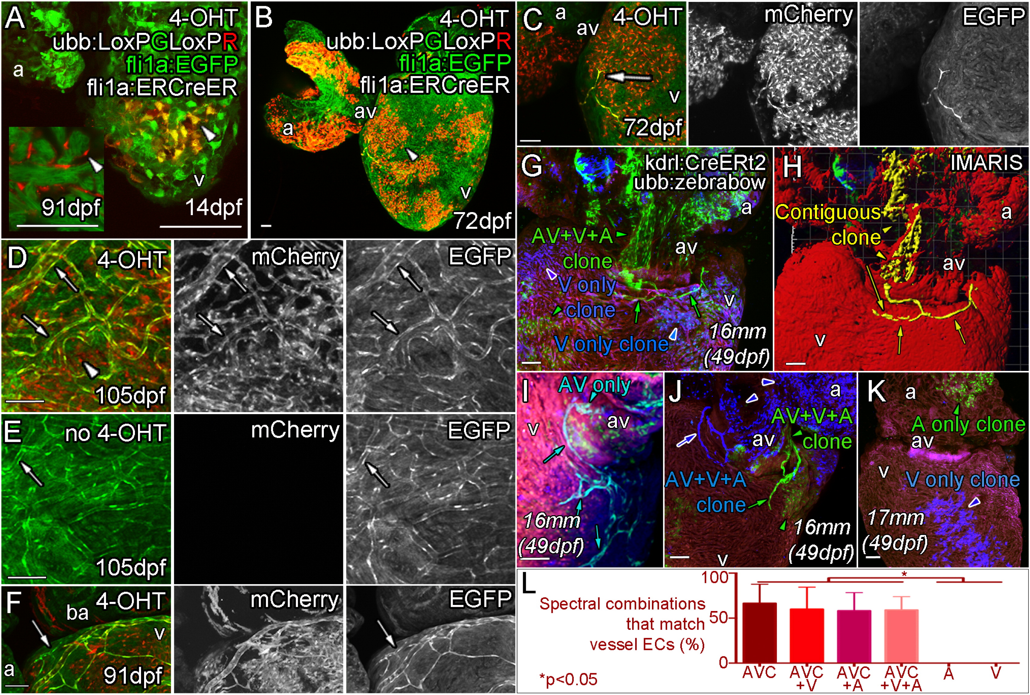
Angiogenic Coronary Endothelial Cells Are Derived from a Subpopulation of the Endocardium
Triple transgenic fish, Tg (ubb:LoxPEGFPLoxPmCherry (ubb:LoxPGLoxPR); fli1aep:ERt2CreERt2 (fli1a:ERCreER); fli1a:EGFP) were treated with 4-OHT during embryogenesis (0–5 dpf) to label the endocardium as mCherry positive clonal patches (arrowheads) and imaged at 14 dpf (A), 72 dpf (B and C), 91 dpf (F) and 105 dpf (D). (E) No 4-OHT control. (A, inset) 91 dpf section with no fli1a:EGFP showing mCherry expression in endocardial cells lining the trabecular myocardium. Emerging endothelial cells (fli1a:EGFP-positive, bright green) are also mCherry-positive (C and D, arrow, labeled endothelial cells; arrowhead, labeled endocardial cells). The majority of subsequently formed vessels are found to be similarly labeled in adult zebrafish treated with 4-OHT as embryos (D, arrows), but not in those that were not exposed to 4-OHT during embryogenesis (E, arrow). Unlabeled vessels (arrowhead) have been observed on hearts with labeled vasculature consistent with the hypothesis that multiple cells give rise to vessels by an angiogenic process (F, arrow). Clonal analysis with multispetral fluorescent proteins (ubb:Zebrabow; ubb:Lox2272-LoxP-RFP-Lox2272-CFP-LoxP-YFP) is used to follow the fate of restrictedly labeled endocardial cells (G–L). Image of the ventral side of the AV canal, with ventricle below and atrium above showing labeling of the endocardial cells as a single green clone (G, green arrowhead), which gives rise to the forming vessel on the ventricle surface (G, green arrows). Part of this green clone is contiguous with the surface endothelial cells, extending through the myocardium to the AV canal and atrium (H, yellow). Analysis of multiple clones, including AV endocardium specific clones (I), shows a clonal relationship between AV endocardial cells and endothelial cells on the surface (G–J). Forming vessels can be derived from more than one clone, but always share the same labeling with the AV canal (J). Non-AV clones are found not to give rise to surface endothelium (K). Quantification of the endocardial spectral combinations found in different heart regions that match those of the vessel endothelium (L, p < 0.05 ANOVA/Tukey, ± SEM). Zebrafish age listed as dpf or italicized total body lengths with equivalent age in brackets when zebrafish were raised under non-standard conditions. AVC, atrioventricular canal; A, atrium; V, ventricle. Scale bars represent 50 µm.
Reprinted from Developmental Cell, 33, Harrison, M.R., Bussmann, J., Huang, Y., Zhao, L., Osorio, A., Burns, C.G., Burns, C.E., Sucov, H.M., Siekmann, A.F., Lien, C.L., Chemokine-guided angiogenesis directs coronary vasculature formation in zebrafish, 442-54, Copyright (2015) with permission from Elsevier. Full text @ Dev. Cell