
Fig. 2
- ID
- ZDB-IMAGE-150708-20
- Publication
- Harrison et al., 2015 - Chemokine-guided angiogenesis directs coronary vasculature formation in zebrafish
- All Figures
- Figures for Harrison et al., 2015
|
Fig. 2
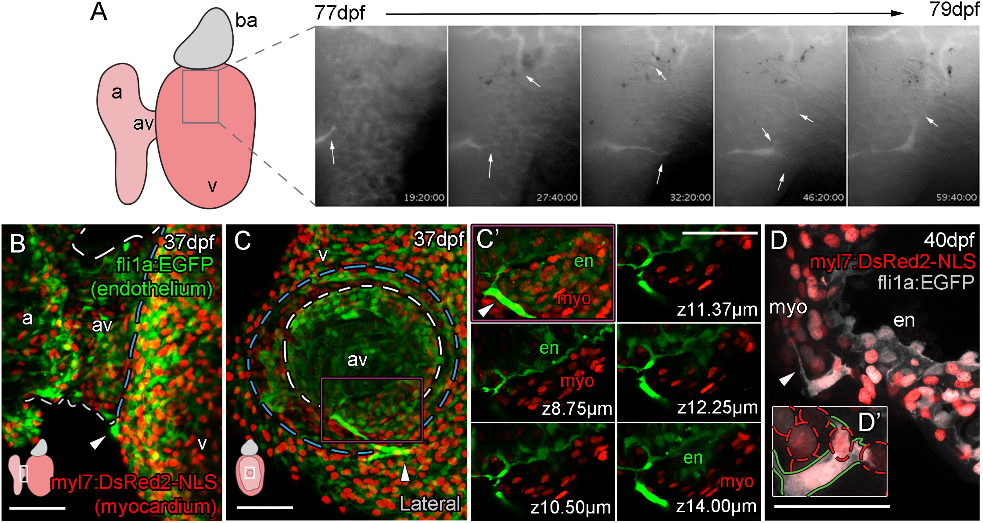
Vessels Are Formed from Angiogenic Sprouts of Cells Emerging from the AV Canal
(A–D′) Live imaging stills show the angiogenic migration of the endothelial cells that migrate over the ventricle surface and form interconnections between forming vessels (A; angiogenic sprouts marked by arrows). The first emerging endothelial cells are visible on the surface of the ventricle at the juncture of the atrium and ventricle (arrowheads, B, ventral view; C, lateral view of the same heart, diagram denotes heart orientation). Cells at this point have a direct connection to the endocardium (C, dashed white line demarks AV canal, blue line marks invagination of the ventricular myocardium; C′, higher magnification of region demarcated by purple box with select individual planes in z, labeled with distance from the first plane of projection). Individual endothelial cells span the myocardium (D; D′, higher magnification with red dashed lines demarcating myocardial nuclei, green line demarcates endothelial cell) and then project filopodia over the ventricle surface (D, arrowhead). Scale bars represent 50 µm.
Reprinted from Developmental Cell, 33, Harrison, M.R., Bussmann, J., Huang, Y., Zhao, L., Osorio, A., Burns, C.G., Burns, C.E., Sucov, H.M., Siekmann, A.F., Lien, C.L., Chemokine-guided angiogenesis directs coronary vasculature formation in zebrafish, 442-54, Copyright (2015) with permission from Elsevier. Full text @ Dev. Cell