
Fig. 1
- ID
- ZDB-IMAGE-150708-19
- Publication
- Harrison et al., 2015 - Chemokine-guided angiogenesis directs coronary vasculature formation in zebrafish
- All Figures
- Figures for Harrison et al., 2015
|
Fig. 1
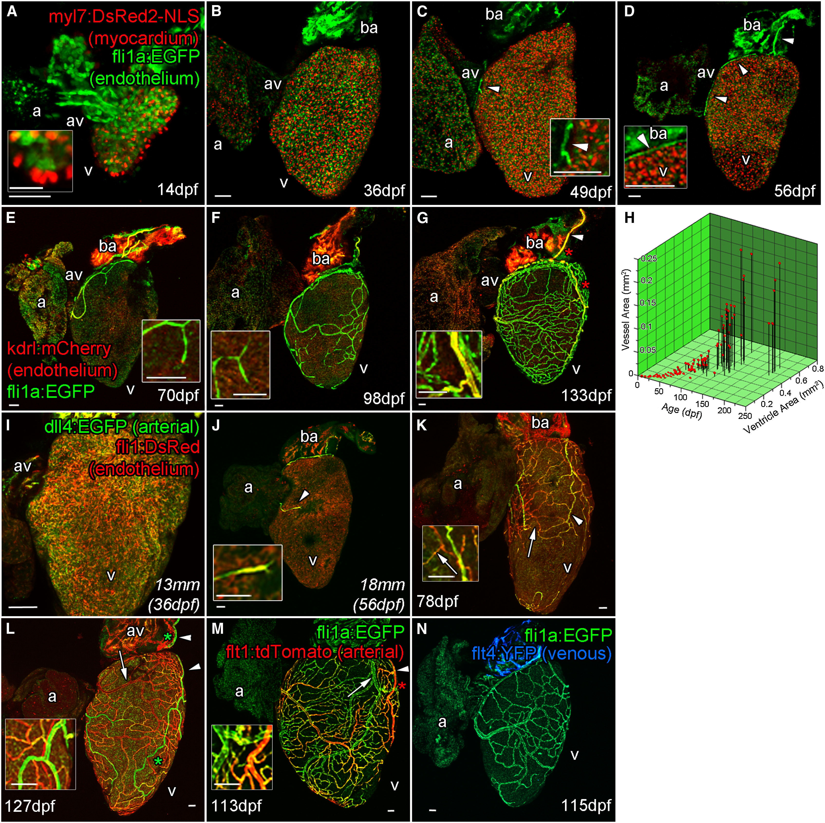
Zebrafish Coronary Vessels Develop during Juvenile Stages and Respond to Increased Heart Size
(A–G) Confocal images of whole larval, juvenile and adult double transgenic hearts, Tg(fli1a:EGFP; myl7:DsRed2-NLS (A–D) and Tg(fli1a:EGFP; kdrl:mCherry) (E–G). flila:EGFP-positive endothelial (endocardial) cells are visible only on the inside of the myocardium (labeled with myl7:DsRed2-NLS) and bulbous arteriosus after hatching (A and inset 14 dpf; and B, 36 dpf). Five to seven weeks later, endothelial cells are found on the surface of the heart (C, 49 dpf, arrowhead and inset), forming a vessel connection between the gills and AV-boundary after 1 month (D, 56 dpf, arrowheads and inset). The major vessel over the bulbous arteriosus expresses high levels of kdrl:mCherry (marked by arrowhead in G). Branches from these early vessels appear to progressively cover and interconnect over the juvenile heart into adulthood; however, the majority express low kdrl:mCherry levels (E, 70 dpf to G, 133 dpf, insets).
(H and I) The emergence and coverage of these vessels is stochastic, but correlates strongly with age and the area of the ventricle (H). Endocardial cells prior to the emergence of vessels express arterial marker dll4:EGFP (I).
(J and K) Emerged endothelial cells on the ventricle surface also have an arterial identity (J, arrowhead and inset), which is selectively downregulated in some vessels as they form (K, arrow).
(L and M) A subset of vessels maintain dll4:EGFP (L, arrowhead) and express flt1:tdTomato confirming they are arteries (M, arrowhead).
(N) Vessels that downregulate arterial makers appear to not progress to full venous identity as flt4:YFP expression is not observed at significant levels. a, atrium; av, AV canal; v, ventricle. Zebrafish age listed as dpf or italicized total body lengths with equivalent age in brackets when zebrafish were raised under non-standard conditions. artery. Scale bars represent 50 µm except for that in (A, inset), which is 10 µm.
Reprinted from Developmental Cell, 33, Harrison, M.R., Bussmann, J., Huang, Y., Zhao, L., Osorio, A., Burns, C.G., Burns, C.E., Sucov, H.M., Siekmann, A.F., Lien, C.L., Chemokine-guided angiogenesis directs coronary vasculature formation in zebrafish, 442-54, Copyright (2015) with permission from Elsevier. Full text @ Dev. Cell