
|
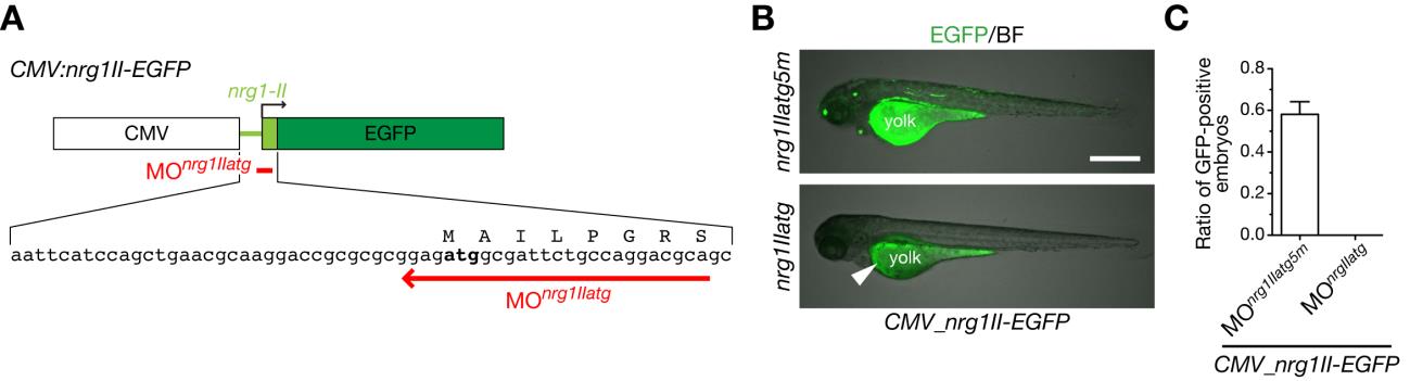
Fig. S7 MOnrg1IIatg specifically suppresses ectopic expression of NRG1-II.
A. A schematic structure of an expression plasmid CMV:nrg1II-EGFP (top), a part of the nucleotide and amino acid sequences encoding 5′ untranslated and coding regions in the first exon of nrg1-II (middle), and the target sequence of MOnrg1IIatg (bottom, red arrow). B. Representative 74-hpf embryos co-injected with CMV:nrg1II-EGFP expression plasmid and MOnrg1IIatg or the control MOnrg1IIatg5m shown in a lateral view. Green fluorescence in yolk of MOnrg1IIatg-injected embryos is autoflurescence (arrowhead). Scale bar, 500 µm. C. Quantification of ratios of GFP-positive embryos co-injected with CMV:nrg1II-EGFP and MOnrg1IIatg or the control MOnrg1IIatg5m at 25 hpf. No GFP-positive embryos were detected for MOnrg1IIatg-injected embryos under a fluorescent dissection microscopy. (mean ± s.e.m.; nrg1IIatg5m, 0.56 ± 0.08, n = 183 embryos by 4 injections, nrg1IIatg, 0.00 ± 0.00, n = 98 embryos by 4 injections).