
|
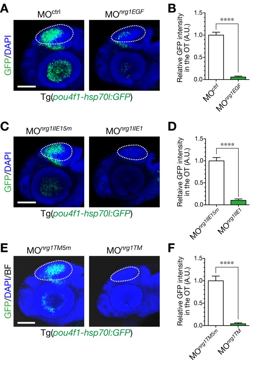
Fig. S5 Defective generation of GFP-expressing neurons in the optic tectum by injection of antisense MOs against nrg1.
A. Knockdown of all isoforms of NRG1 by injection of MOnrg1EGF. Representative embryos injected with MOnrg1EGF (right) or the control MOctrl (left) shown in a lateral view at 48 hpf. Dotted circle, optic tectum (OT). Scale bar, 100 µm. B. Quantification of pou4fl-hsp70l:GFP intensity in the OT for the experiment shown in A (mean ± s.e.m.; ****P < 0.0001, unpaired t test; n = 6–7). C. Impaired neurogenesis in a representative MOnrg1IIE1-injected Tg(pou4f1-hsp70l:GFP) embryo (right) compared to the control MOnrg1IIE15m (5 nucleotides-mismatched control)-injected embryo (left) at 50 hpf. Scale bar, 100 µm. D. Quantification of pou4f1-hsp70l:GFP intensity in the OT for the experiment shown in C (mean ± s.e.m.; ****P < 0.0001; n = 8 per group). E. Knockdown of membrane-bound isoforms of NRG1 by injection of MOnrg1TM. Representative embryos injected with MOnrg1TM (right) or the control, MOnrg1TM5m (left) shown in a lateral view at 53 hpf. Dotted circle, OT. Scale bar, 100 µm. F. Quantification of pou4f1-hsp70l:GFP intensity in the OT for the experiment shown in E (mean ± s.e.m.; ****P < 0.0001, unpaired t test; n = 6 per group).