
|
Fig. S9
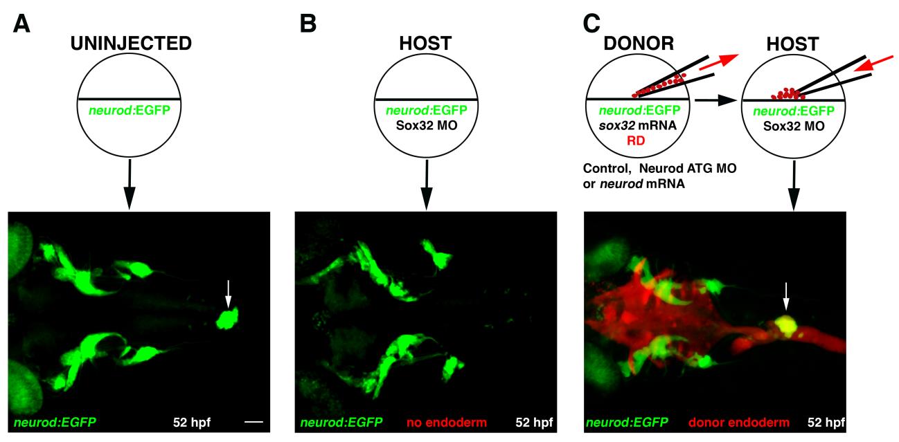
Cell transplantation approach.Tg(neurod:EGFP) embryos were used to follow the fate of transplanted cells in chimeric embryos. (A) Confocal image (merged z-stack) of an uninjected 52 hpf Tg(neurod:EGFP) embryo; arrow indicates EGFP expression in the pancreas. (B) Sox32 morphant embryos, which lack endoderm, were used as hosts. (C) Tg(neurod:EGFP) donor embryos were injected with rhodamine dextran (RD; red) lineage tracer and sox32 mRNA. Donors were additionally injected with nothing, Neurod ATG MO or neurod mRNA, to generate control, Neurod morphant or Neurod overexpressing donor cells, respectively. Donor cells were transplanted into Sox32 morphant hosts. A confocal image is shown of a chimeric embryo with a fully reconstituted donor-derived endoderm (red) expressing EGFP-positive endocrine cells (arrow). Anterior to left in all specimens. White scale bar=50 µm.
Reprinted from Developmental Biology, 402(1), Dalgin, G., Prince, V.E., Differential levels of Neurod establish zebrafish endocrine pancreas cell fates, 81-97, Copyright (2015) with permission from Elsevier. Full text @ Dev. Biol.