
|
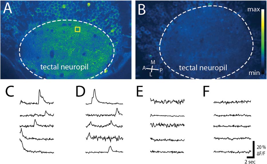
Fig. 2
TTX reduced both spontaneous and induced activity in the tectal neuropil. (A,B) Images of the tectal neuropil in HuC:GCaMP5 fish at 4 dpf when injected (A) with a control (vehicle alone) or (B) TTX. 250 frames of each movie were stacked for maximum intensity over time and psuedocolored with an imageJ Look Up Table (Green Fire Blue) where yellow shows the most intense activity over a blue background. (C, D) Spontaneous activity, as well as activity induced by the initiation of the 488 nm laser, was observed in the tectal neuropil in control cases at (C) 4 dpf and (D) 5 dpf. (E, F) When injected with TTX at 3.5 dpf no spiking activity was observed in the neuropil at (E) 4 dpf or (F) 5 dpf. Yellow box in (A) shows an example of a 25 × 25 pixel region chosen for activity analysis. A; anterior, M; medial, P; posterior, L; lateral.