
|
Fig. 1
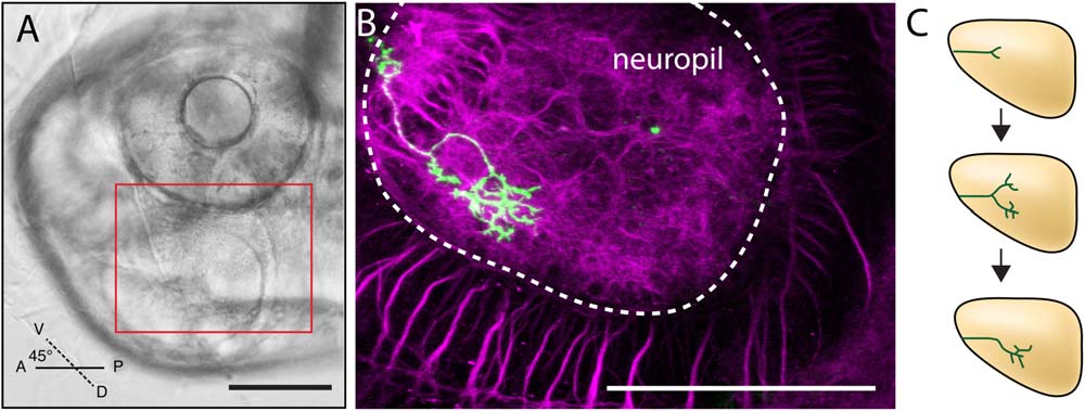
Imaging RGC growth on the tectum. (A) Brightfield view of a 54 hpf, PTU-treated zebrafish mounted in agar, dorsal side down and at a 45° angle to position the tectum close to the lens for inverted microscopy. The red rectangle marks the optic tectum and the approximate area imaged during pathfinding. (B) Confocal image of a fixed and immunostained whole mount after imaging at 5.5 dpf. An axon (mGFP, green) has arborized in the neuropil (dotted white outline). Only the right tectal neuropil is visible, with the arbor coming from a RGC in the left eye (out of frame). Anti-acetylated tubulin (magenta) allowed the architecture of the area to be visualized. (C) Schematic summarizing the mechanism identified by Simpson et al. (2013) for guidance by biased branching. Branches are extended and retracted dynamically during pathfinding, with more branches extending toward the target at any given time. Branches that decrease the distance to the target could become the primary axon shaft when other branches are retracted. Sequential rounds of branching refine the direction of travel, and this iterative process continues until the target is reached. Scale bars are 100 µm. A; anterior, D; dorsal, P; posterior, V; ventral.