
|
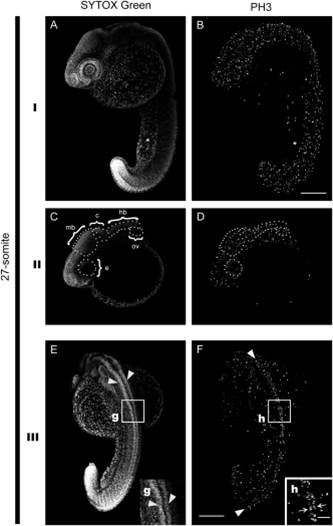
Fig. 12
Proliferation patterns in 27-somite stage embryos. I: Left-side views. II: Partial projections were most superficial slices were omitted to highlight internal patterns. III: Dorsal views. The embryo shows abundant PH3 positive nuclei in the main axis as compared to the yolk cell. The dorsal region of the neural tube (white arrowheads) shows mitotic nuclei arranged in two rows along antero–posterior length of the embryo as in the previous stage analyzed. (g, h inserts): show an amplification to highlight the structure and proliferation patterns in the neural tube. e, eye; mb, midbrain; c, cerebellum; hb, hindbrain; ov, otic vesicle. 200 µm scale bar in B. 50 µm scale bar in h insert.