
Fig. 4
- ID
- ZDB-IMAGE-150609-10
- Genes
- Publication
- Love et al., 2015 - Rest represses maturation within migrating facial branchiomotor neurons
- All Figures
- Figures for Love et al., 2015
|
Fig. 4
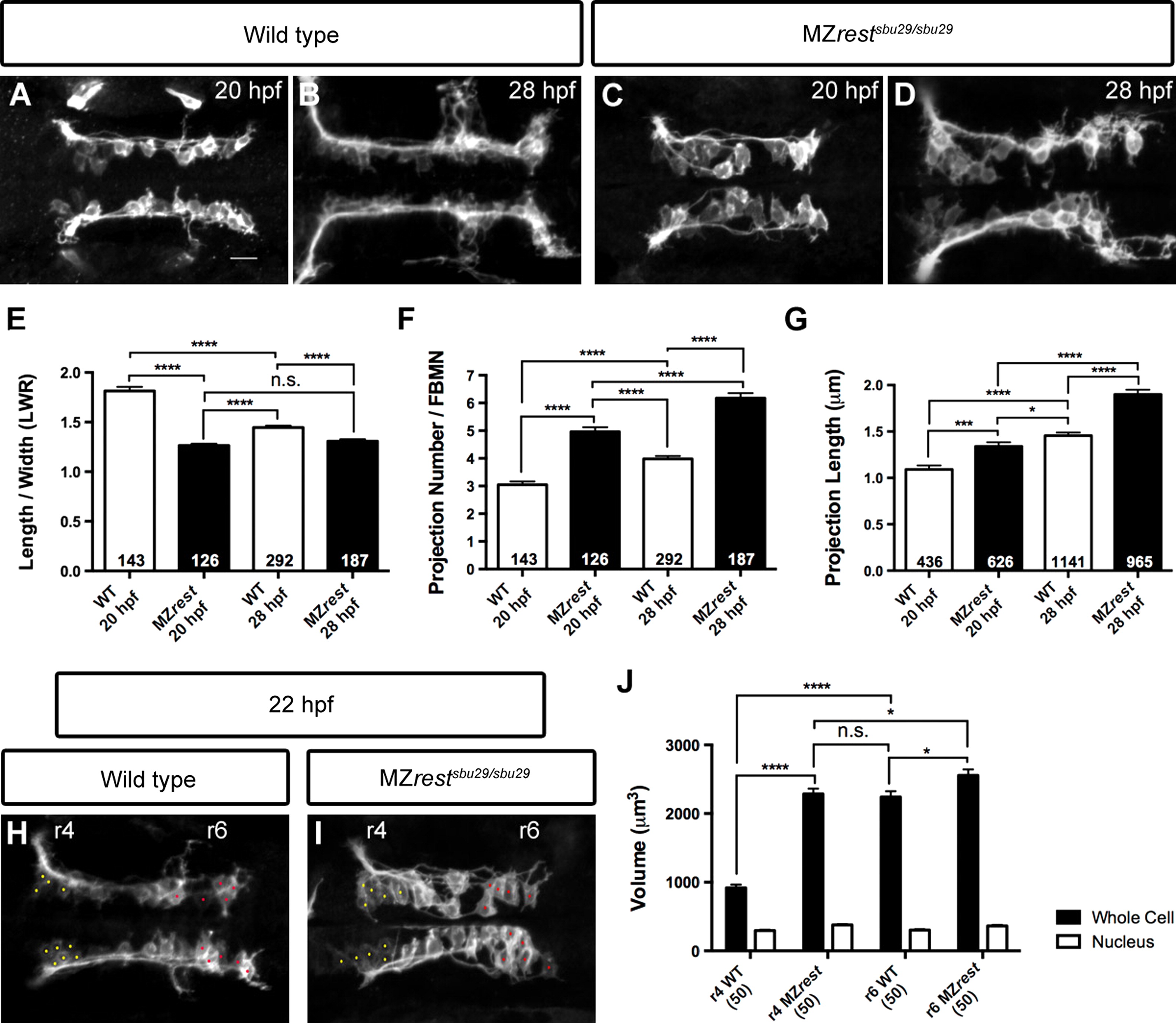
Morphological analysis reveals precocious maturation of Rest-deficient FBMNs. (A–G) Analysis of FBMNs at 20 and 28 hpf in Tg(zCREST1:membRFP) wild type embryos (A and B) and MZrestsbu29/sbu29 embryos (C and D). Average measurements from 5 embryos are shown for wild type and MZrestsbu29/sbu29 (E–G) embryos at each stage. The wild type length-to-width ratio decreases from 20 to 28 hpf, as neurons become rounder; by contrast MZrestsbu29/sbu29 FBMNs are round at both 20 and 28 hpf (E). Both number and length of protrusions increase over time in wild type and MZrestsbu29/sbu29 embryos (F and G). MZrestsbu29/sbu29 FBMNs have significantly more and longer protrusions than wild type FBMNs. (H–J) Volume measurements of FBMNs at 22 hpf. Whole cell and nuclear volume were measured for 10 “immature” neurons in r4 (yellow dots) versus 10 “mature” neurons in r6 (red dots) for 5 wild type (H) and 5 MZrestsbu29/sbu29 (I) embryos. Wild type FBMN whole cell volume increases significantly as neurons migrate from r4 to r6; MZrestsbu29/sbu29 FBMN whole cell volume does not alter as neurons migrate from r4 to r6 and is similar to that of wild type neurons in r6. Nuclear volume does not change significantly. Mean±SEM; *P<0.05; ***P<0.001, ****P<0.0001, n.s.=not significant.
Reprinted from Developmental Biology, 401(2), Love, C.E., Prince, V.E., Rest represses maturation within migrating facial branchiomotor neurons, 220-35, Copyright (2015) with permission from Elsevier. Full text @ Dev. Biol.