
|
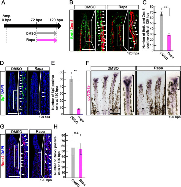
Fig. 6
Rapamycin treatment inhibits proliferation and differentiation of osteoblasts after 72 hpa. (A) Scheme of rapamycin treatment between 72 and 120 hpa. (B, C) BrdU and Zns-5 double-stained fin sections and quantification of BrdU and Zns-5 double-positive osteoblasts. The number of double-positive osteoblasts was significantly reduced by rapamycin treatment at 120 hpa. **p < 0.01 by Student’s t-test. Error bars represent the standard error of 4 independent experiments. (D, E) Sp7-stained fin sections and quantification of Sp7-positive osteoblasts. The number of Sp7-positive osteoblasts was significantly reduced by rapamycin treatment at 120 hpa. DAPI fluorescent signal (blue) indicates the presence of nuclei. **p < 0.01 by Student’s t-test. Error bars represent the standard error of 4 independent experiments. Scale bars: 100 µm. (F) Expression of col10a1a was examined by in situ hybridization at 120 hpa (n = 4). Rapamycin treatment decreased col10a1a expression (brackets in F) at 120 hpa. Dashed lines indicate the amputation planes. Scale bars: 200 µm. (G, H) Runx2-stained fin sections and quantification of Runx2-positive cells. The number of Runx2-positive cells was not affected by rapamycin treatment at 120 hpa. Error bars represent the standard error of 4 independent experiments. Scale bars: 100 µm.