
|
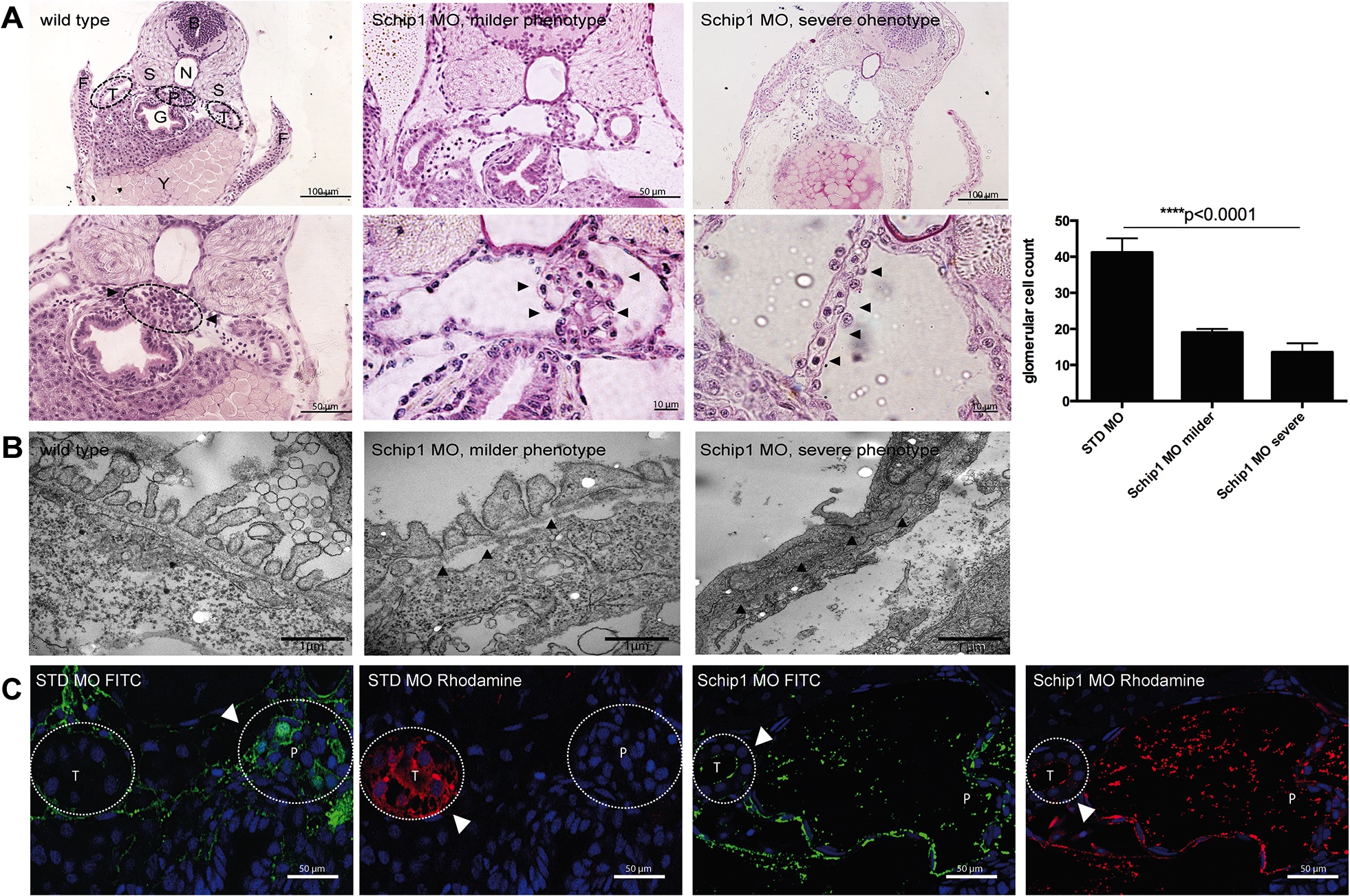
Fig. 4 Schip1 inactivation in zebrafish causes distortion of podocyte foot processes, podocytopenia and leakage of the filtration barrier.
(A) Brightfield images of PAS stained histological sections showing dilated Bowman’s space and distortion of proper podocyte structures (arrowheads) in Schip1 morphant zebrafish with milder (second panel) and more severe phenotype (third panel). Higher magnifications (size bars) shown in lower panel, including a cell nuclei quantification graph. (B) Electron microscopic analyses of zebrafish morphants show effaced podocyte foot processes with various degrees of damage (middle, arrowheads and right). However, slit diaphragms are consistently present (right, arrowheads). (C) Dye filtration assay in control and Schip1 zebrafish morphants. Rhodamine conjugated 10kDa is freely filtered into the tubuli of both control and morphant fish, whereas FITC labeled 500kDa dye remains in the pronephros in control embryos. In Schip1 morphants 500kDa dye accumulates in the Bowman’s space and also leaks into the tubuli. STD-standard control morpholino, F-fins, T-nephric tubuli, S-somites, B-brain, N-notochord, P-pronephros, G-gut, Y-yolk.