
|
Fig. 6
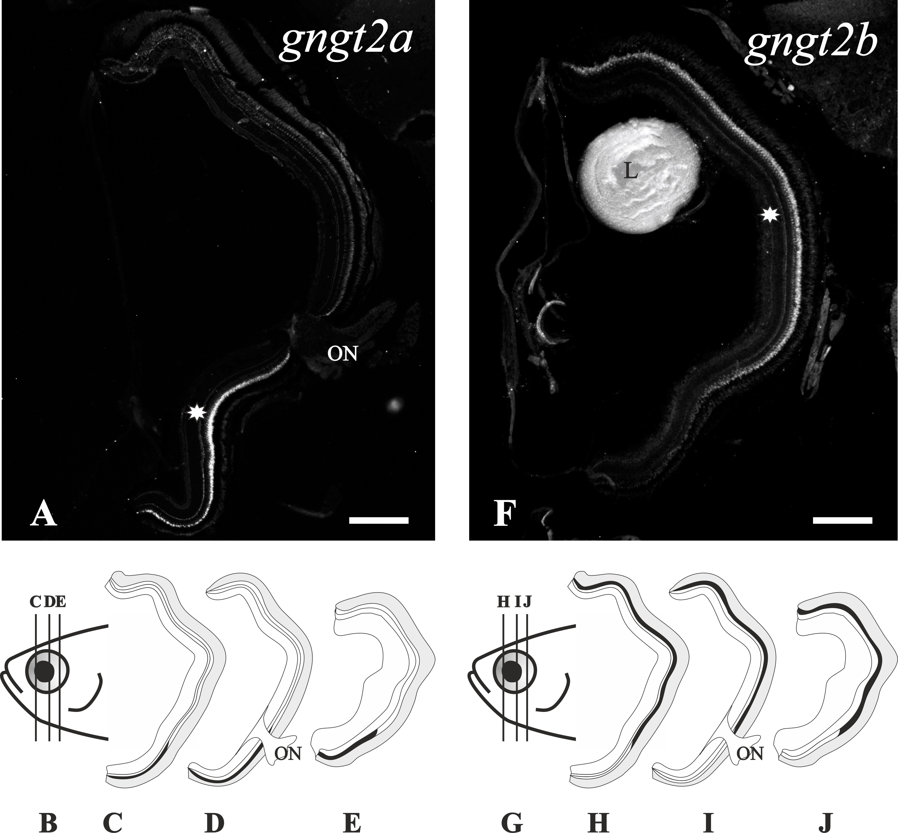
Compartmentalisation of the gngt2a and gngt2b cone-specific paralogs in the adult retina.
The upper part shows two photomicrographs from radial sections of adult zebrafish eyes. The inner side is to the right and the dorsal side to the top. In them, expression patterns (asterisks) can be observed in the ventral retina for the gngt2a gene (A) and in the dorsal and medial retina for the gngt2b gene (F). At the bottom are two drawings of a zebrafish adult head in a lateral view (B, G), pointing out the level where the sections were taken for the schematic drawings in panels C, D and E for gngt2a, and panels H, I and J for gngt2b. L; lens, ON; optic nerve. Scale bars: 200 µm.