
|
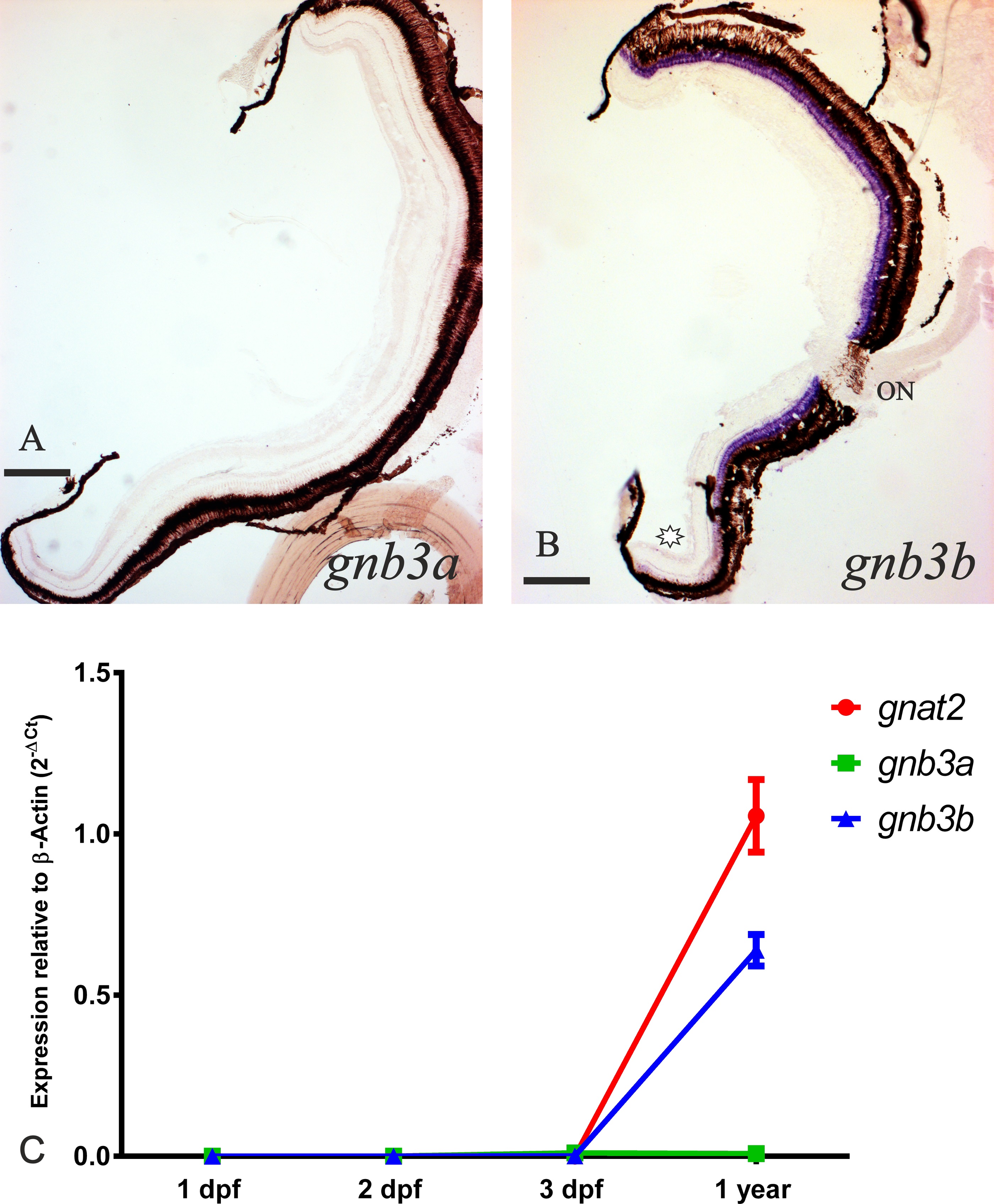
Fig. 5
Expression of the gnb3 paralogs retained in zebrafish after 3R.
The upper part of the figure shows two photomicrographs from radial sections of adult zebrafish eyes. The inner side is to the right and the dorsal side to the top. No mRNA could be detected for the gnb3a gene (A). Expression of the gnb3b gene (in purple) was observed in the dorsal and medial retina (B), while the ventral retina lacks mRNA (asterisk). The black-brown tissue surrounding the retina corresponds to the pigmentary epithelium. ON; optic nerve. Scale bars: 200 µm. At the bottom of the figure there is a graph comparing the cone-specific gnat2, gnb3a and gnb3b expression levels between 1 dpf, 2 dpf, 3 dpf zebrafish and eyes of 1 year old individuals in relation to the β-actin housekeeping gene using the 2-ΔCt method (C). Note the increment in the expression of gnat2 and gnb3b in the adult stage while the similar amount of mRNA for the gnb3a gene present in the 3 dpf embryos and an adult eye.