
|
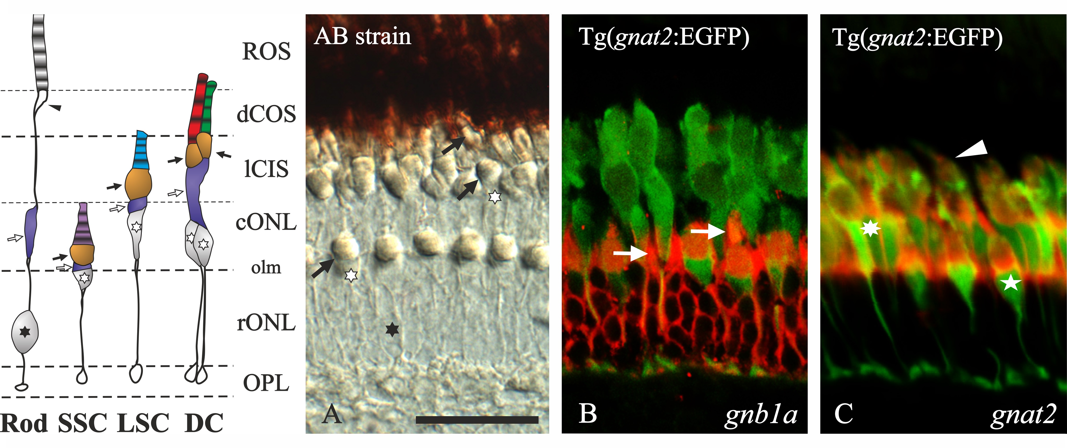
Fig. 2
Adult zebrafish outer retina; structure and probe specificity.
The schematic diagram of the outer retina to the left is based on Raymond and Barthel, 2004 [36] and a Nomarski contrast photomicrograph from a radial section of an adult AB zebrafish retina (A). All photoreceptors have a terminal in the outer plexiform layer (OPL). The outer nuclear layer (ONL) is subdivided by the outer limiting membrane (olm) into a sublayer where the rods have their nuclei (black asterisk: rONL) and a more external sublayer where the cones have their nuclei (empty asterisks: cONL). Note that the short single cones (SSC) have their nuclei in the rONL while their oil droplets (black arrows) and outer segments (OS) are in the cONL. The rods’ myoids (empty arrows) are embedded in the cONL. The long cone inner segment sublayer (lCIS) includes the oil droplets and myoids of the double cones (DC) and the long single cones (LSC) and part of the LSC OS. The outermost part of the retina (top) is covered by the pigmentary epithelium where the DC OS (dCOS) and the rods’ ellipsoid (arrowhead) and outer segments (ROS) are embedded. B and C are confocal photomicrographs of in situ hybridisation experiments performed using Tg(gnat2:EGFP) zebrafish, which expresses EGFP in all cones. Specific Fast Red staining in rods for gnb1a (B) and cones for gnat2 (C) can be observed. Arrows in B point to the rod´s myoids that in some cases could be mistaken as SSC. In C, the arrowhead points to a DC, the asterisk to a LSC and the star to a SSC. Scale bar in A represents 25 µm.