
|
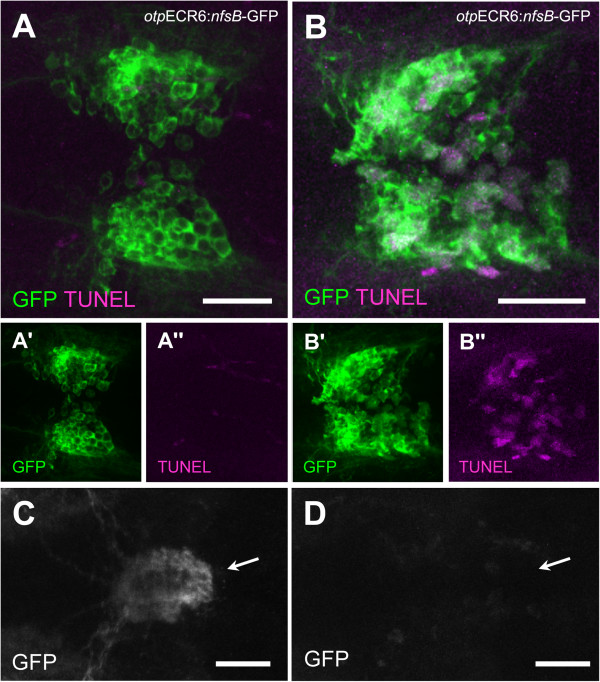
Fig. 6
Nitroreductase-expressing cells inTg(otpECR6-E1b:nfsb-GFP)larvae display a reduction in the number of projections to the pituitary and an increase in apoptotic bodies when exposed to Mtz. A, TUNEL staining of the preoptic GFP expression domain in an untreated larva. B, TUNEL staining of the preoptic GFP expression domain in an Mtz-treated larva. C, Pituitary bundles of GFP-stained fibers in a control larva (arrow). D, The pituitary location in an Mtz-treated larva (arrow). Note that in the control larva, the GFP fibers reach the pituitary and no apoptosis is detected, but in the Mtz-treated larva, the pituitary innervation is disrupted and apoptosis is apparent in preoptic cell bodies. Scale bars: 50 µm.