
IMAGE
Fig. 5
- ID
- ZDB-IMAGE-150601-134
- Genes
- Antibodies
- Source
- Figures for Gutierrez-Triana et al., 2014
Image
|
Figure Caption
Fig. 5
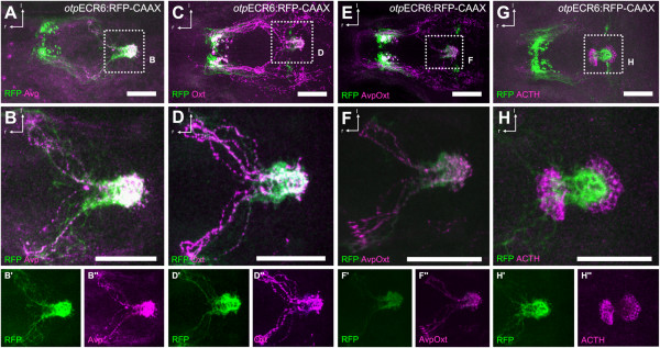
A group of cells labeled by the activity of theotpECR6 enhancer project to the pituitary. A-D, Costaining of RFP with Avp (A) or Oxt (C) as typical hypophysiotropic cell types shows a high degree of overlap of fibers reaching the pituitary (magnified views in B and D). E, Costaining of RFP with Avp and Oxt combined (magnified view in F). G, Costaining with ACTH as a pituitary marker confirms the dense RFP bundles as part of the pituitary (magnified view in H). Abbreviations: r, rostral; l, lateral. Scale bars: 100 µm.
Figure Data
Acknowledgments
This image is the copyrighted work of the attributed author or publisher, and
ZFIN has permission only to display this image to its users.
Additional permissions should be obtained from the applicable author or publisher of the image.
Full text @ BMC Dev. Biol.