
Fig. 4
|
Fig. 4
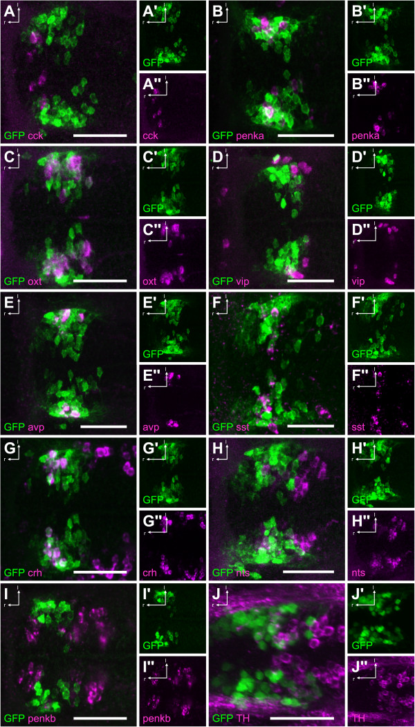
NPO cells involved in the HPA axis are labeled in theTg(otpECR6-E1b:mmGFP)transgenic line. A, There is no overlap of GFP with in situ stained cck+ cells. B, There is some overlap of GFP with penka+ cells. C, There is extensive overlap of GFP with oxt+ cells. D, There is no overlap of GFP with vip+ cells. E, There is a high degree of overlap of GFP with avp+ cells. F, There is some overlap of GFP with sst1.1+ cells. G, There is a high degree of overlap of GFP with crh + cells. H, There is some overlap of GFP with nts+ cells. I, There is no overlap of GFP with penkb + cells. J, There is no overlap of GFP with immunostained TH+ cells. Abbreviations: r, rostral; l, lateral. Scale bars: 50 µm.