
|
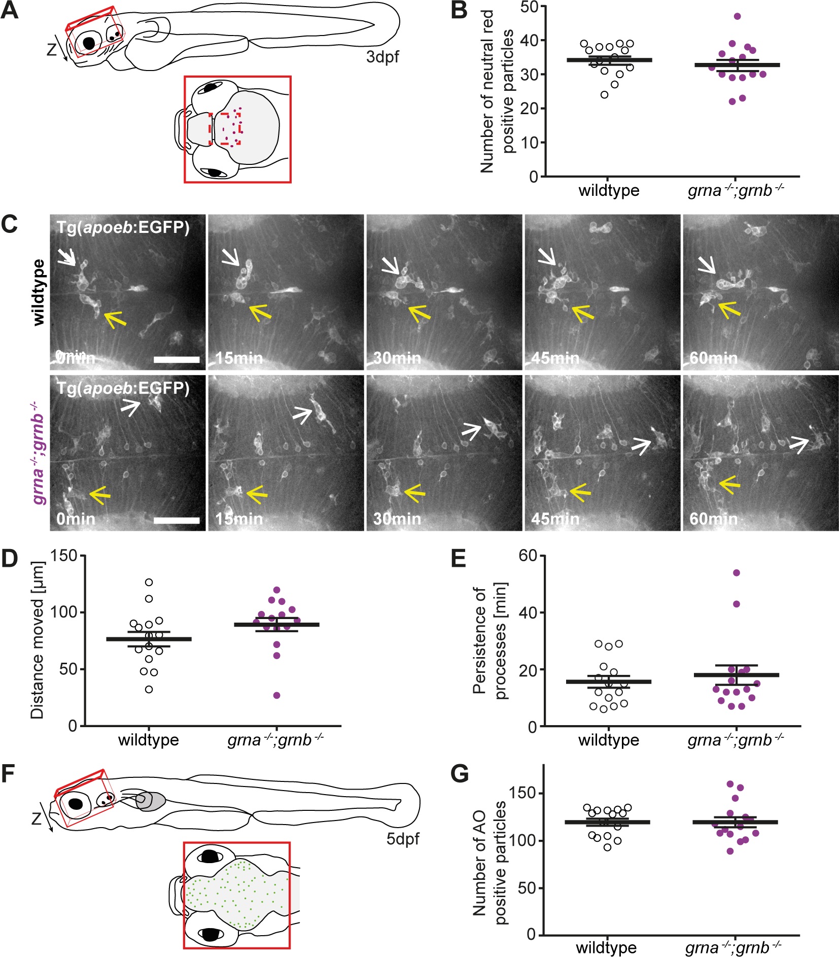
Fig. 4 No microgliosis and neurodegeneration in grna-/-;grnb-/- mutants.
A: Schematic illustration of a zebrafish larvae at 3dpf (lateral view) and a detail of the region (red line), dorsal view, imaged for the analysis of neutral red positive particles (B). The dashed red line marks the area that was imaged in the time lapse recordings of microglia (C). B: The number of neutral red positive particle in the region illustrated in A (Z-stack) is unchanged in wildtype and grna-/-;grnb-/- mutants. n = 15. S.E.M. Mann-Whitney test (two-tailed). p = 0.2884. C-E: Microglia in Tg(apoeb:lynEGFP) grna-/-;grnb-/- mutants and wildtype larvae at 3dpf are indistinguishable. C: Still images of the time lapse recordings in the optic tectum recorded by spinning disk confocal microscopy. Two microglia cells marked in each genotype by a white and yellow arrow. Dorsal view. Anterior to the left. n = 3. Scale bar: 50µm. Recording time: 60min. 1frame/min. D: The distance microglia move within one hour in grna-/-;grnb-/- mutants and wildtype larvae. Quantification of n = 3x5 randomly selected microglia from the time lapse recordings shown in C. S.E.M. Mann-Whitney test (two-tailed). p = 0.0671. E: Processes in the grna-/-;grnb-/- mutants and wildtype larvae persist for same durations. Quantification of n = 3x5 randomly selected processes from the time lapse recordings shown in C. S.E.M. Mann-Whitney test (two-tailed). p = 0.8296. F: Schematic illustration of a zebrafish larvae at 5dpf (lateral view) and a detail of the region, dorsal view, imaged for the analysis of acridine orange (AO) positive cells. G: The number of acridine orange positive cells in the region illustrated in C (Z-stack) is unchanged in wildtype and grna-/-;grnb-/- mutants. n = 15. S.E.M. Mann-Whitney test (two-tailed). p = 0.69.