
|
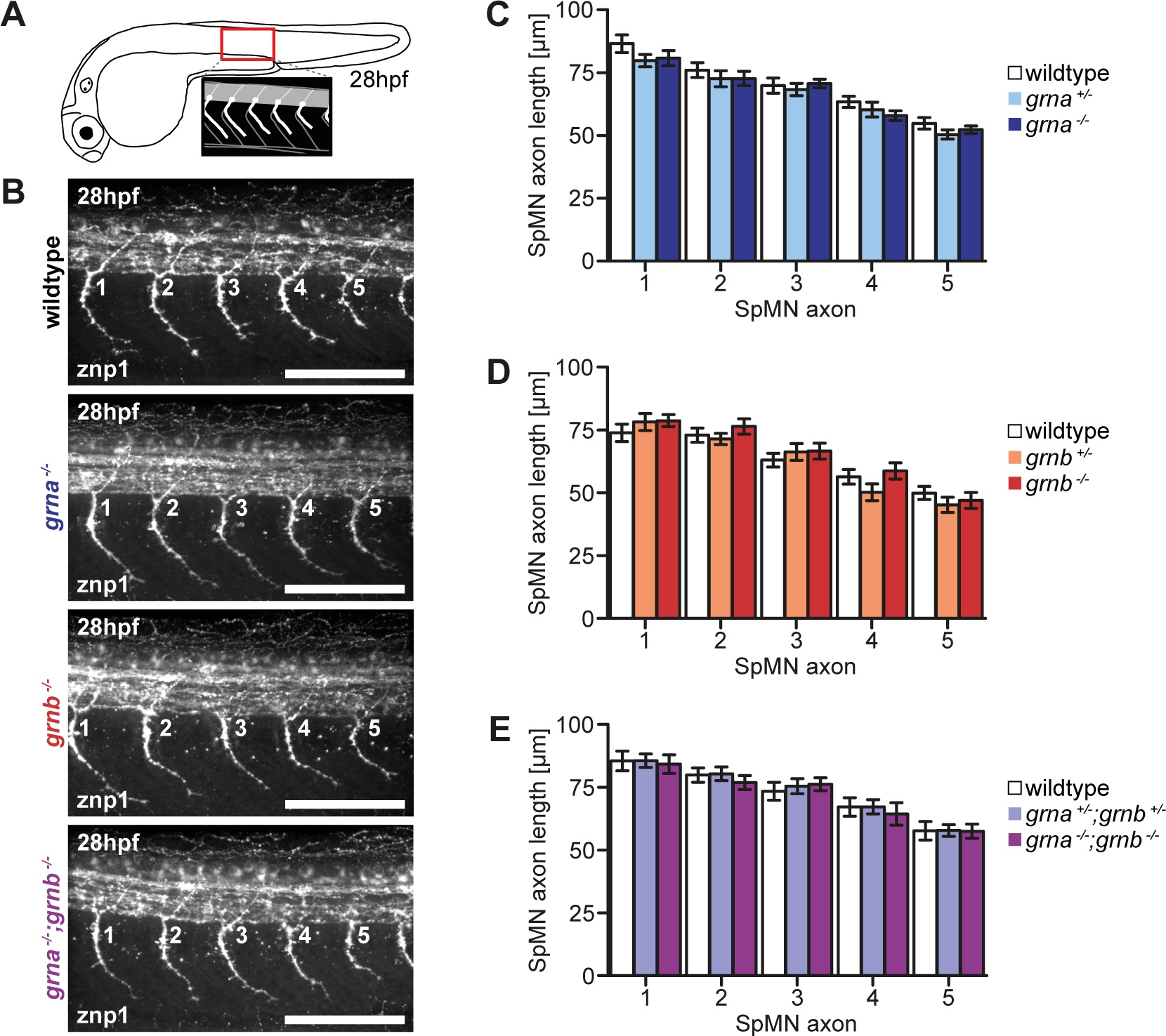
Fig. 2 No SpMN axonopathy in Grna and Grnb single and double KOs.
A: Schematic illustration of a zebrafish embryo at 28hpf (lateral view) and a detail of the region above the end of the yolk extension imaged for the analysis of SpMN axons (lateral view). B: In Grna and Grnb single and double KOs the SpMN axons show no extended branching. Whole-mount immunofluorescence staining of 28hpf embryos with znp1 antibody. The 5 SpMN axons above the end of the yolk extension are shown. Images taken by spinning disk confocal microscopy. Anterior to the left. Lateral view. Orthogonal projections. Scale bar: 100µm. C-E: Quantification of the SpMN axon length in homozygous and heterozygous Grna and Grnb single and double KOs and wildtype siblings. The SpMN axon length of the 5 SpMN axons (1–5) above the end of the yolk extension is measured from the exit point of the spinal cord to the tip of the growth cone. C: Homozygous and heterozygous Grna KOs and wildtype siblings. n = 30. D: Homozygous and heterozygous Grnb KOs and wildtype siblings. n = 30. E: Homozygous and heterozygous Grna and Grnb KOs and wildtype siblings. n = 25. S.E.M. Two-way ANOVA. Bonferroni post-test. All non-significant (n.s.).