
|
Fig. S5
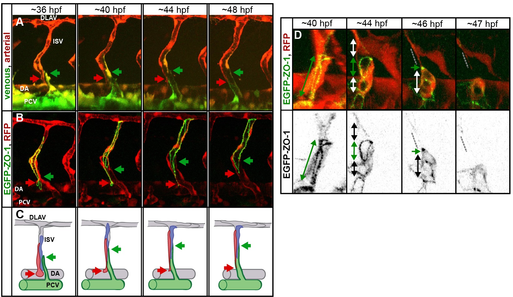
Pruning in the ISVs.
(A) Stills of a time-lapse movie showing segmental vein formation in a transgenic embryo Tg(fliep:GFF)ubs4; Tg(UAS:Kaede)rk7. Arterial cells are marked in red, venous cells are green (photoconverted Kaede, colors are inverted for better visualization). A new venous sprout (green arrow) grows out of the PCV towards a segmental vessel (red arrow). The venous sprout connects to the ISV. At the same time, the ISV segment connected to the aorta narrows and eventually detaches as the cells migrate up to contribute to the dorsal part of the ISV. See also S12 Movie. (B) Still images of the segmental vein formation in a transgenic embryo Tg(fliep:GFF)ubs4; Tg(UAS:mRFP);Tg(UAS:EGFP-ZO-1)ubs5. A single cell is expressing the EGFP-ZO-1 (green). The cell belongs to the ISV and is anchored in the DA (red arrow points to the ring-like junction). When the venous sprout connects (green arrow), the green cell moves up the ISV. The junctional ring gradually reduces in size to a spot, and the cell eventually detaches from the aorta and migrates up the ISV. See also S13 Movie. (C) A cellular model of pruning during the segmental vein formation. Arteries are grey, and venous cells are green. Red and blue cells are initially connected to the DA. As the venous sprout attaches to the ISV, the arterial cells detach from the DA and move up to contribute to the dorsal part of the ISV. (D) Stills from a time-lapse movie showing multicellular-to-unicellular tube transformation during pruning in an ISV in a transgenic embryo Tg(fliep:GFF)ubs4; Tg(UAS:mRFP);Tg(UAS:EGFP-ZO-1)ubs5. Key steps of pruning are shown, corresponding to the model in Fig 6. Green arrows mark multicellular contacts (cell–cell junction length), white arrows mark transcellular lumen, and grey dotted lines mark unicellular fragments without lumen.