
|
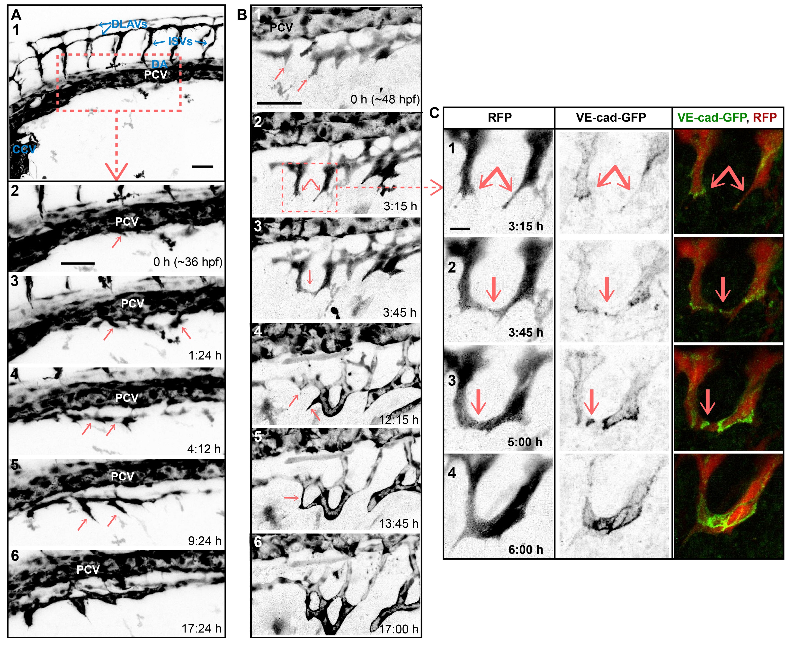
Fig. S1
SIV outgrowth involves single cell assembly and angiogenic sprout fusion.
Stills of time-lapse movies showing outgrowth of the SIVs between ~36 and ~60 hpf in transgenic embryos: Tg(fli1a:EGFP) in A and Tg(fliep:GFF)ubs3,(UAS:mRFP), (5xUAS:cdh5-EGFP)ubs12 in B and C. (A) SIV emerges from the PCV at ~36 hpf (1–2). Single cells sprout ventrally from the PCV (2–3, arrows). The cells that contact each other (4, arrows) often lose contact with the PCV. A single, nonlumenized primary SIV forms and produces ventral angiogenic sprouts (5, arrows) and eventually lumenizes (6). PCV, posterior cardinal vein; DA, dorsal aorta; CCV, common cardinal vein; ISV, intersegmental vessel; DLAV, dorsal longitudinal anastomotic vessel. See also S3 Movie. (B) Multiple angiogenic sprouts originating from the primary SIV connect to each other (arrows) before (1–3) and after (4–6) lumen inflation, forming a net of vascular loops. (C) Close-up of SIV sprout fusion including the VE-cad-EGFP channel, showing new contact formation (2, arrow marks a spot of junctions), contact expansion (3, a ring of junctions), and transformation of the new branch into a multicellular tube (lines of junctions in 4). See also S4 Movie. Scale bars: 50 µm (A and B) and 10 µm (C).