
|
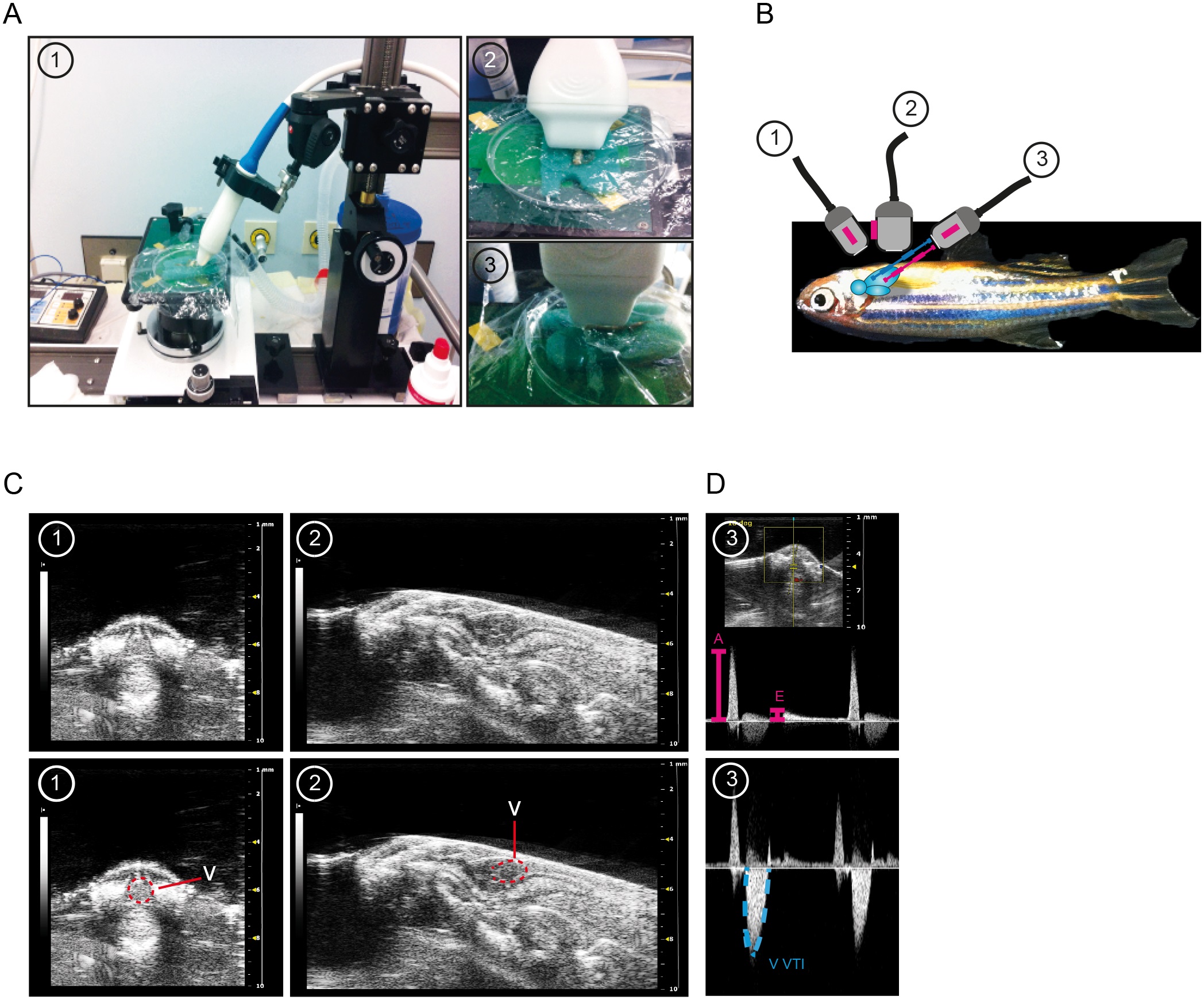
Fig. 1
Three plane echocardiography to assess heart function in adult zebrafish.
(A) Overview of experimental setting and illustration of transducer positioning to image short axis view (SAX) (1), abdominal-cranial axis (ACX) for pulsed-wave Doppler (PWD) recordings (2), and long axis view (LAX) (3). (B) Lateral view of dorsally positioned adult zebrafish with three defined transducer positions illustrated. The red line indicates direction of ultrasound beam for PWD acquisition for cardiac inflow velocity imaging and the blue line indicates ultrasound beam direction for acquisition of cardiac outflow velocities. (C) Representative images of SAX view (1) and LAX view (2) acquisition (upper row). In the lower row the ventricle is outlined in red in (1) and (2). (D) PWD images derived from the ACX view (3) with the upper image showing representative signals attained from the AV-valve region with clearly visible positive A- and E- waves (in pink) and the bottom image displaying one representative V VTI PWD signal obtained from the bulbus arteriosus region. PWD, pulsed-wave Doppler.