
Image
|
Figure Caption
Fig. 2
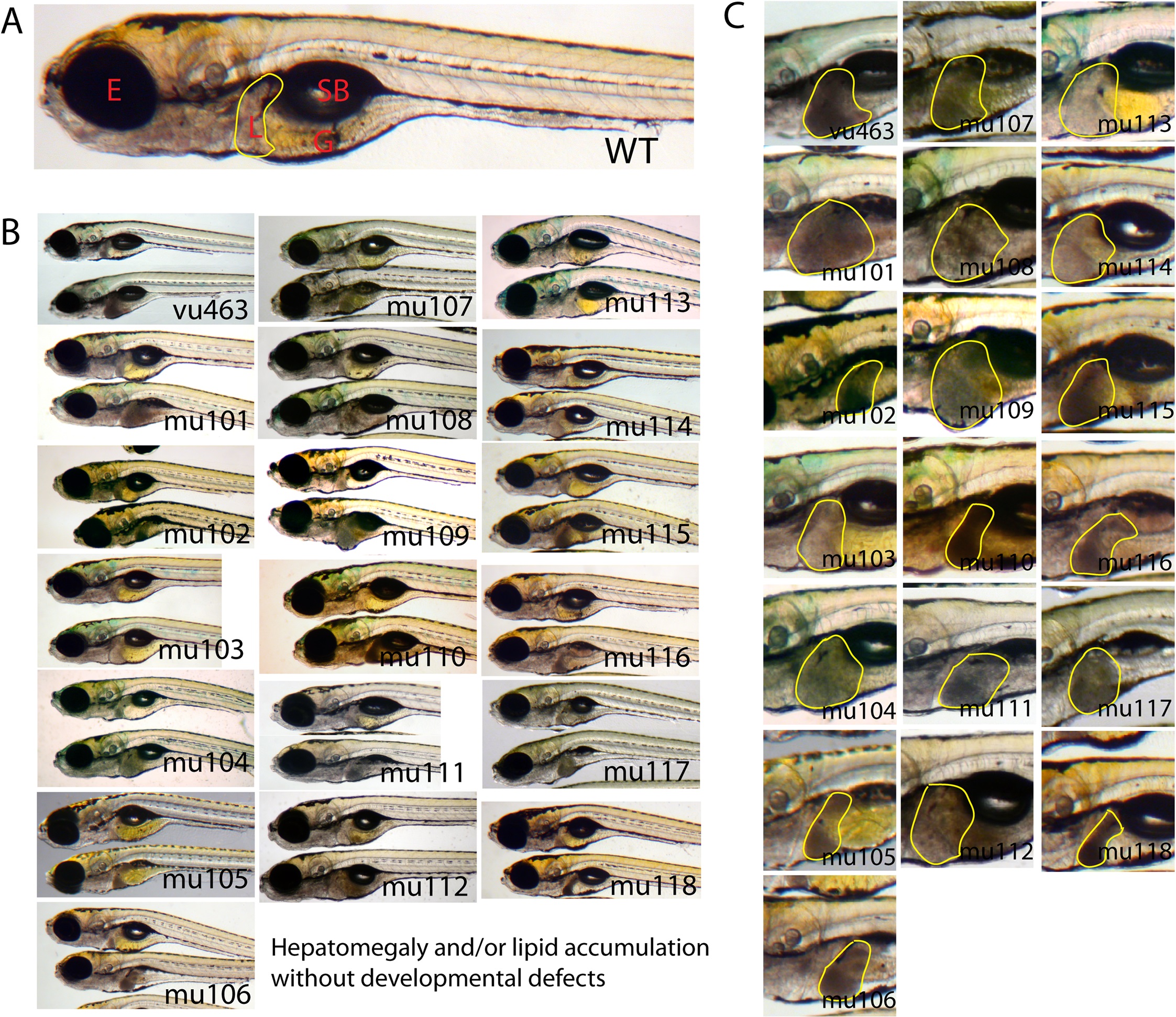
Zebrafish mutants exhibit post-developmental liver defects without developmental phenotypes.
(A) A wild type control at 7 dpf. Livers are outlined with yellow line. (B) Control siblings are on the top and homozygous mutants are on the bottom of each panel. (C) Magnified images of livers (outlined with yellow) in homozygous mutants. E = eye, L = liver, G = gut and SB = swim bladder. “mu”, stands for MUSC, followed by a number that designates the unique mutants identified in the screen. n>100 per each mutant.
Figure Data
Acknowledgments
This image is the copyrighted work of the attributed author or publisher, and
ZFIN has permission only to display this image to its users.
Additional permissions should be obtained from the applicable author or publisher of the image.
Full text @ PLoS One