
Image
|
Figure Caption
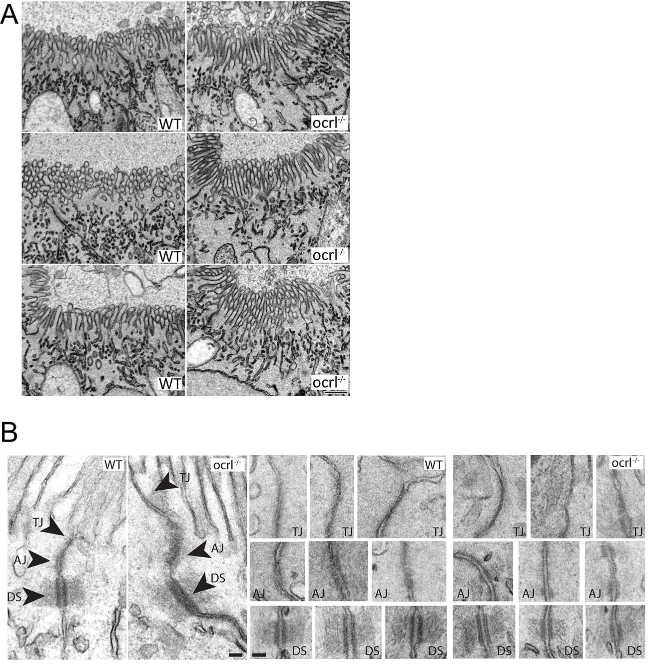
Fig. S6 Brush border and intercellular junctions of ocrl-/- zebrafish pronephric cells.
A. Block face scanning electron microscopy images of microvilli at the apical brush border of pronephric tubule cells of wild type and ocrl-/- embryos (72hpf). B. Transmission electron microscopy images of intercellular junctions between pronephric cells of wild type and ocrl-/- embryos (72hpf). AJ = adherent junctions, TJ = tight junctions, DS = desmosomes. Scale bars represent 0.5 µm (A) and 100 nm (B).
Acknowledgments
This image is the copyrighted work of the attributed author or publisher, and
ZFIN has permission only to display this image to its users.
Additional permissions should be obtained from the applicable author or publisher of the image.
Full text @ PLoS Genet.