
|
Fig. 3
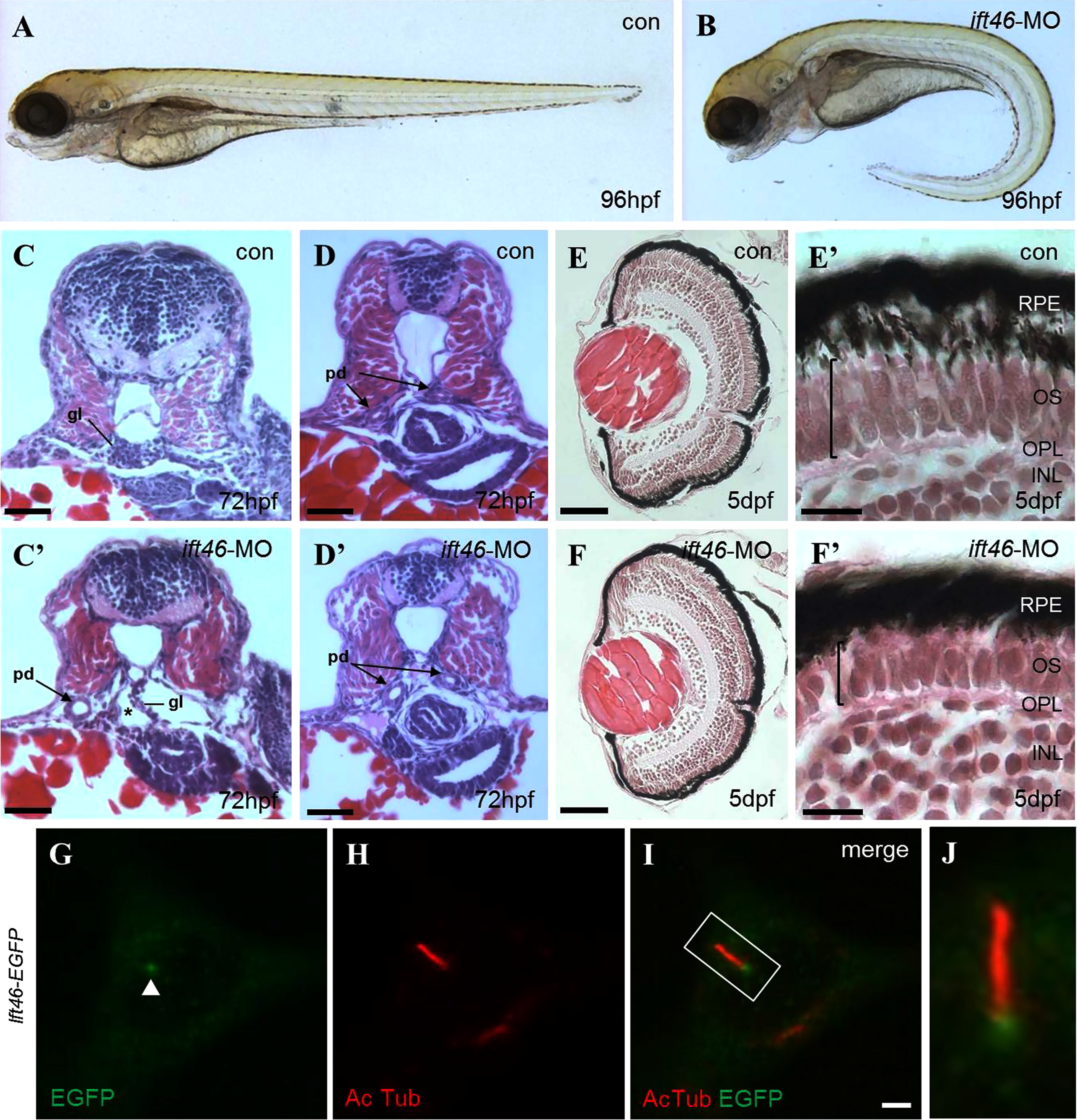
ift46 morphants develop phenotypes associated with ciliary dysfunction. (A) Wild-type embryos show normal morphology. (B) ift46 morphants are characterized by curved body axis, the development of pronephric cyst and pericardiac edema at 96 hpf. (C–D) Cross-sections of wild-type zebrafish at the glomerular–tubular region of pronephros at 72 hpf show normal morphology of pronephric glomeruli and ducts. (Có–Dó) ift46 morphant embryos display a grossly distended cyst (asterisk) in place of glomeruli (gl) and dilated pronephric ducts (pd) (arrow). Scale bar, 50 µm. (E–F′) Histological sections of the eyes of wild-type and ift46 morphants at 5 dpf. The retina in wild-type control (E and E′) has normal laminated structures, including retinal pigment epithelium (RPE), outer segment (OS), outer plexiform layer (OPL) and inner nuclear layer (INL). (E) and (F) scale bar, 200 µm. The outer segment (OS) (bracket) is thinner in ift46 morphants (F′) compared to the control (E′). Scale bar, 100 µm. (G–J) The EGFP-tagged ift46 (green) is localized to the base of primary cilia (red) in hTERT-RPE1 cells. Merged imaged are shown in (I). Arrowhead indicates the base of cilia. Scale bar, 3 µm. (J) Higher magnification view of the boxed area in (I).
Reprinted from Developmental Biology, 400(2), Lee, M.S., Hwang, K.S., Oh, H.W., Ji-Ae, K., Kim, H.T., Cho, H.S., Lee, J.J., Yeong Ko, J., Choi, J.H., Jeong, Y.M., You, K.H., Kim, J., Park, D.S., Nam, K.H., Aizawa, S., Kiyonari, H., Shioi, G., Park, J.H., Zhou, W., Kim, N.S., Kim, C.H., IFT46 plays an essential role in cilia development, 248-57, Copyright (2015) with permission from Elsevier. Full text @ Dev. Biol.