
|
Fig. S3
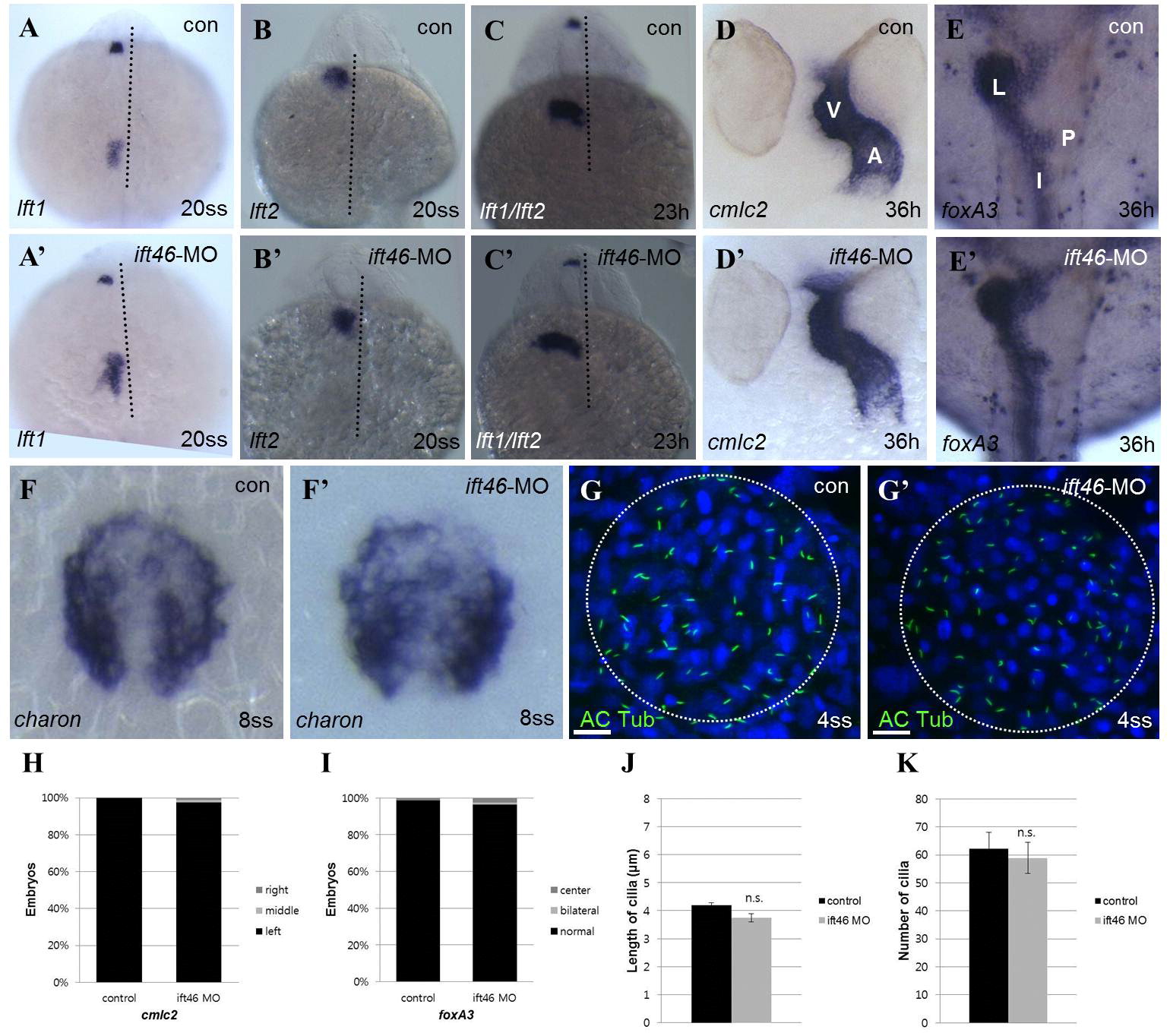
The organ laterality defect is not disrupted in zebrafish ift46 morphants. (A–C′) The ift46 morphant embryos have no left–right patterning defect visualized by whole-mount in situ hybridization for lft1 and lft2 (D–E′). The ift46 morphants does not have any abnormal heart looping and gut laterality with whole-mount in situ hybridization for cmlc and foxA3. (A, artery; I, intestine; L, liver; P, pancreas; V, vein). (F–F′) Expression of charon in Kupfferós vesicle in the control and the ift46 morphants are comparable. (G–G′) Visualization of cilia with immunostaining of acetylated α-tubulin in the Kupfferós vesicle in both control and the ift46 morphants at 6-somite stage. Scale bar, 20 µm. (H and I) Organ asymmetry scored by cmlc2 (Control (n=56) and ift46 morphants (n=85)) and foxA3 (Control (n=68) and ift46 morphants (n=81)) expression. (J and K) Quantification of the number and length of the KV cilia shows no statistical differences between the control and the ift46 morphant embryos.
Reprinted from Developmental Biology, 400(2), Lee, M.S., Hwang, K.S., Oh, H.W., Ji-Ae, K., Kim, H.T., Cho, H.S., Lee, J.J., Yeong Ko, J., Choi, J.H., Jeong, Y.M., You, K.H., Kim, J., Park, D.S., Nam, K.H., Aizawa, S., Kiyonari, H., Shioi, G., Park, J.H., Zhou, W., Kim, N.S., Kim, C.H., IFT46 plays an essential role in cilia development, 248-57, Copyright (2015) with permission from Elsevier. Full text @ Dev. Biol.