
Fig. 1
- ID
- ZDB-IMAGE-150506-41
- Genes
- Publication
- Moreno et al., 2014 - Spinal neurons require Islet1 for subtype-specific differentiation of electrical excitability
- All Figures
- Figures for Moreno et al., 2014
|
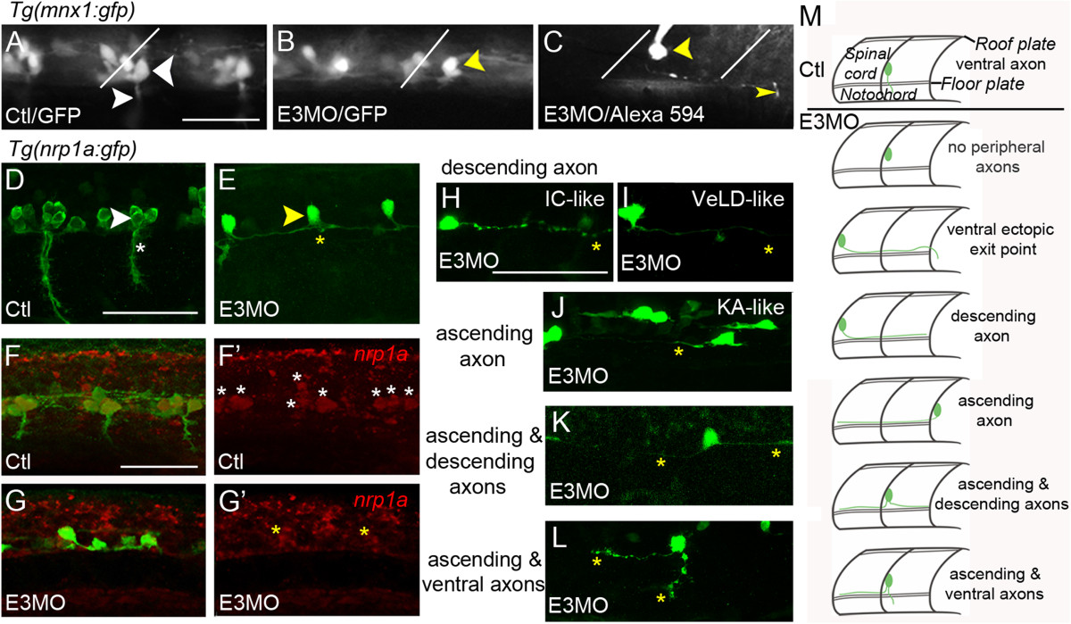
Fig. 1
Caudal primary motor neuron-like neurons have abnormal axonal trajectories that resemble those of interneurons. In all figures, unless indicated otherwise, images present lateral views of embryos (rostral left, dorsal up). (A) In control (Ctl) Tg(mnx1:gfp)ml2 22 to 26 hours post-fertilization (hpf) embryos, primary motor neurons (PMNs) express green fluorescent protein (GFP). The caudal PMN (CaP; large white arrowhead) projects its peripheral axon (small white arrowhead) ventrally. White lines (A, B and C) indicate somite boundaries. (B) In Tg(mnx1:gfp)ml2 E3 morphants, GFP+ cells persist in the ventral spinal cord. GFP+ CaP-like cells (yellow arrowhead) have somas in CaP positions but project axons centrally rather than peripherally. (C) In another Tg(mnx1:gfp)ml2 E3 morphant, a GFP+ CaP-like cell (yellow arrowhead), filled with Alexa 594, has an axon that extends caudally to exit in the neighboring hemisegment (thin yellow arrowhead). (D-L) (D) In uninjected Tg(nrp1a:gfp)js12 embryos, CaP (white arrowhead) has its soma immediately dorsal to the motor axon exit point (white asterisk). (E) E3 morphants have few GFP+ ventral neurons. A CaP-like cell (yellow arrowhead) lacks a peripheral axon. (F, F′) GFP+ PMNs of control Tg(nrp1a:gfp)js12 embryos (F′, white asterisks) express nrp1a. (G, G′) Following Islet1 knock-down, few nrp1a/GFP+ (G′, yellow asterisks) neurons are present. (H-L) In E3 morphants, many GFP+ neurons have axons that bypass normal exit points and extend centrally either caudally (H and I), rostrally (J) or in both directions (K). Occasionally, a GFP+ CaP-like cell extends a peripheral as well as a central axon (L). (M) The top cartoon depicts control CaP axon morphology, and the six lower cartoons exemplify the range of CaP-like axonal phenotypes revealed by either dye filling or confocal analysis of Tg(nrp1a:gfp)js12 E3 morphants. Scale bars = 50 µm in A (for A to C), D (for D and E), F (for F to G′) and H (for H to L). IC, ipsilateral commissural; KA, Kolmer-Agduhr; VeLD, ventral lateral.