
Fig. 6
|
Fig. 6
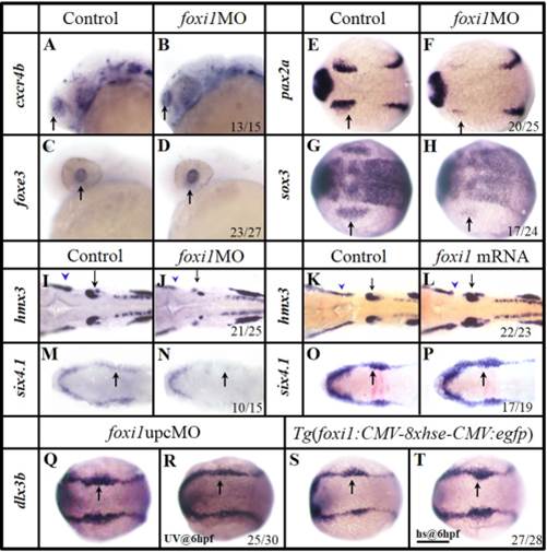
Foxi1 is specifically involved in the OEPD development. A–H: In foxi1 morphants, the olfactory (B) and lens (D) expression are unchanged, compared with their corresponding controls (A,C). The otic primordial pax2a (F) and epibranchial primordial sox3 (H) are severely reduced, compared with their corresponding controls (E, G). I,J: Smaller lateral line placode (blue arrowhead) and otic vesicle (black arrow) develop in foxi1MO embryos (J), compared with the control (I). K,L: The otic vesicles (black arrow) and lateral line placode (blue arrowhead) are enlarged when excess foxi1 mRNA (L) is present, compared with the control (K). M,N: foxi1MO injection results in the compromised caudal PPR six4.1. O,P: Over expression of foxi1 by mRNA injection enhances the OEPD dlx3b. Q,R: Expression of dlx3b is reduced caudally (in OEPD, black arrow) when Foxi1 function is knocked down by an upcMO activated by UV light at 6 hpf. S,T: Over expression of foxi1 by heat shocking a transgenic line Tg(gfp:hse:foxi1) at 6 hpf expands the caudal dlx3b expression (black arrow). A–D: Side views with anterior to the left; E–T: Dorsal views with anterior to the left. A–D and I–J: 24 hpf; E–F:12 hpf; G–H and M–T: 11 hpf; K,L: 26 hpf. Scale bar = 200 µm.